Genetic Abnormalities and Mutation
Help Questions
MCAT Biology › Genetic Abnormalities and Mutation
Which of the following mutations might lead to the formation of a recessive allele?
I. Silent
II. Frameshift
III. Nonsense
IV. Missense
II, III, and IV
I and IV
I, II, III, and IV
II and III
Explanation
Recessive alleles are often created by mutated versions of functional genes that encode broken/nonfunctional proteins. The question is thus asking us which of the mutations is likely to result in a nonfunctional or abnormal protein.
Silent mutations are types of mutations that result in the insertion of the same amino acid due to the degeneracy of the genetic code and, therefore, will not cause any noticeable change in the protein. Essentially, even organisms with the mutation will still show the dominant allele.
Frameshift mutations change the reading frame used for translation and oftentimes result in premature stop codons. Nonsense mutations are mutations that specifically result in premature stop codons and generally result in nonfunctional proteins. Missense mutations lead to the insertion of a different amino acid at the normal site. This can lead to serious problems for the functionality of the protein, particularly if the mutation is present in the active site or another area that is highly conserved throughout evolution.
Human chromosomes are divided into two arms, a long q arm and a short p arm. A karyotype is the organization of a human cell’s total genetic complement. A typical karyotype is generated by ordering chromosome 1 to chromosome 23 in order of decreasing size.
When viewing a karyotype, it can often become apparent that changes in chromosome number, arrangement, or structure are present. Among the most common genetic changes are Robertsonian translocations, involving transposition of chromosomal material between long arms of certain chromosomes to form one derivative chromosome. Chromosomes 14 and 21, for example, often undergo a Robertsonian translocation, as below.

A karyotype of this individual for chromosomes 14 and 21 would thus appear as follows:

Though an individual with aberrations such as a Robertsonian translocation may be phenotypically normal, they can generate gametes through meiosis that have atypical organizations of chromosomes, resulting in recurrent fetal abnormalities or miscarriages.
During meiosis, gametes result from the isolation of one chromosome from each of a human’s homologous 23 pairs. In Figure 2, der(14,21) is treated as a homologous pair to either 14 or 21. An egg, then, could be formed that had a normal chromosome 21 and the der(14,21) instead of the normal chromosome 14. If a sperm fertilizes this egg, and the sperm has a normal chromosome 14 and 21, what will result?
Trisomy
Pleiotropy
Triploidy
Polyploidy
Unbalanced translocation
Explanation
Since the maternal oocyte would have two copies of chromosome 21, the normal 21 and the derivative chromosome, the normal addition of the sperm would lead to a zygote with a total of three copies of chromosome 21, or trisomy 21. Polyploidy is a tempting choice, but polyploid is a state characterized by extra copies of multiple chromosomes, as might be expected from more than one sperm fertilizing an egg.
Cryptosporidium is a genus of gastrointestinal parasite that infects the intestinal epithelium of mammals. Cryptosporidium is water-borne, and is an apicomplexan parasite. This phylum also includes Plasmodium, Babesia, and Toxoplasma.
Apicomplexans are unique due to their apicoplast, an apical organelle that helps penetrate mammalian epithelium. In the case of cryptosporidium, there is an interaction between the surface proteins of mammalian epithelial tissue and those of the apical portion of the cryptosporidium infective stage, or oocyst. A scientist is conducting an experiment to test the hypothesis that the oocyst secretes a peptide compound that neutralizes intestinal defense cells. These defense cells are resident in the intestinal epithelium, and defend the tissue by phagocytizing the oocysts.
She sets up the following experiment:
As the neutralizing compound was believed to be secreted by the oocyst, the scientist collected oocysts onto growth media. The oocysts were grown among intestinal epithelial cells, and then the media was collected. The media was then added to another plate where Toxoplasma gondii was growing with intestinal epithelial cells. A second plate of Toxoplasma gondii was grown with the same type of intestinal epithelium, but no oocyst-sourced media was added.
You are conducting a study of an isolated tribe in New Guinea, and you find that there is widespread resistance to cryptosporidium infection. Upon examination, you find that the resistance is caused by a change in one nucleotide pair in a gene on chromosome 13. What kind of genetic change does this likely reflect?
Single nucleotide polymorphism (SNP)
Chromosomal change
Copy number variant
Genomic imprinting
Fragile X
Explanation
This would be an example of a single nucleotide polymorphism. A fairly common variant is some change at a single base pair in human DNA. It is possible that this base pair change results in a modified protein that functions just as well as the normal protein in most conditions.
Some of these changes may, simply by chance, be better at resisting disease. When exposed to stress, such as a disease epidemic, it is possible this variant of normal becomes the most widespread genotype. This is especially noticeable in an isolated population, like a tribe in New Guinea. Note that not all single nucleotide polymorphisms will have positive effects, and in some cases can cause disease instead of resistance.
An mRNA sequence is supposed to read UAUGGA, but a mutation replaces the second uracil base with guanine. What is the most specific term for this type of mutation?
Nonsense
Missense
Frame-shift
Deletion
Insertion
Explanation
This mutation replaced UAU (a coding codon) with UAG (a stop codon). The most descriptive term for this kind of replacement is a nonsense mutation.
Note that an easy way to remember the stop codons is UGA ("u" get away), UAA ("u" are away), and UAG ("u" are gone).
Which answer choice correctly shows a frameshift mutation?
CGGTGAATAGGC 
CGGTGAATAGGC 
CGGTGAATAGGC 
CGGTGAATAGGC 
CGGTGAATAGGC 
Explanation
A framshift mutation is when an extra base is inserted into (or deleted from) a strand of DNA. When the DNA is transcribed, every codon downstream from the insertion is changed because the reading frame will be different. This will be the case for any mutations that affect a sequence of nucleotides that is not a multiple of three, for example a five-nucleotide deletion. Such mutations affect the three-nucleotide groupings for all codons following the mutation, and often result in a premature stop codon.
The correct answer is CGGTGAATAGGC 
Note that the reading frame does not change in the following 3-nucleotide insertion. Though there is an extra codon, the downstream sequence is not affected.
CGG-TGA-ATA-GGC 
Type II diabetes results from defective pancreatic beta cells and increased insulin resistance, indicating that peripheral tissues (such as skeletal muscle) do not properly respond to insulin.
Mouse models have been developed to model type II diabetes. In addition to global mutations, tissue-specific mutations can be used to delete genes of interest in precise regions of the body. A group of investigators is interested in characterizing the role of the gene Dia in the onset of diabetes.
Four groups of male mice are compared. Group A is a control group, group B has a global deletion of Dia, group C has a beta cell-specific Dia mutation, and group D has a skeletal muscle-specific Dia mutation.
In order to measure the ability of these mice to respond to a glucose challenge, the mice are fasted overnight. Following the fast, their blood glucose levels are measured (in mg/dL). The mice are then injected with two grams of glucose, and blood glucose levels are measured at 30, 60, 90, and 120 minutes post-injection.
| | 0 min | 30 min | 60 min | 90 min | 120 min | | | ------------ | ---------- | ---------- | ---------- | ----------- | --- | | Group A | 80 | 150 | 120 | 90 | 80 | | Group B | 90 | 220 | 180 | 160 | 140 | | Group C | 100 | 260 | 190 | 150 | 135 | | Group D | 75 | 145 | 110 | 90 | 75 |
Based on the data, what role does Dia play in insulin regulation?
Promote insulin production
Promote insulin sensitivity
Decrease insulin production
Decrease insulin sensitivity
Initiate beta cell deterioration
Explanation
The beta cell-specific Dia mutation in group C causes results similar to the global Dia mutation in group B. From this, we can conclude that Dia is functioning within the beta cells. Essentially, deleting Dia from the beta-cells is equivalent to deleting it from the entire body. Additionally, loss of Dia in skeletal muscle in group D seems to have no phenotypic effect, indicating that Dia is not necessary in skeletal muscle.
Pancreatic beta cells are responsible for insulin production, while skeletal muscle plays a significant role in insulin sensitivity. Since loss of Dia in beta cells leads to high blood glucose, we can conclude that the role of Dia is in the promoting the production of insulin, and that the gene plays no role in insulin sensitivity.
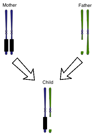
The concept of genomic imprinting is important in human genetics. In genomic imprinting, a certain region of DNA is only expressed by one of the two chromosomes that make up a typical homologous pair. In healthy individuals, genomic imprinting results in the silencing of genes in a certain section of the maternal chromosome 15. The DNA in this part of the chromosome is "turned off" by the addition of methyl groups to the DNA molecule. Healthy people will thus only have expression of this section of chromosome 15 from paternally-derived DNA.
The two classic human diseases that illustrate defects in genomic imprinting are Prader-Willi and Angelman Syndromes. In Prader-Willi Syndrome, the section of paternal chromosome 15 that is usually expressed is disrupted, such as by a chromosomal deletion. In Angelman Syndrome, maternal genes in this section are deleted, while paternal genes are silenced. Prader-Willi Syndrome is thus closely linked to paternal inheritance, while Angelman Syndrome is linked to maternal inheritance.
Figure 1 shows the chromosome 15 homologous pair for a child with Prader-Willi Syndrome. The parental chromosomes are also shown. The genes on the mother’s chromosomes are silenced normally, as represented by the black boxes. At once, there is also a chromosomal deletion on one of the paternal chromosomes. The result is that the child does not have any genes expressed that are normally found on that region of this chromosome.
In addition to the chromosomal deletion on chromosome 15 in the passage, the father is found to have another gene with a mutation, which adds a stop codon prematurely in the base pair sequence. This mutation is best described as a __________.
nonsense mutation
conservative missense mutation
frameshift mutation
silent mutation
non-conservative missense mutation
Explanation
The best answer is a nonsense mutation, which is defined as a point mutation that gives rise to an early stop codon, thus truncating any protein products prematurely. These are typically devastating mutations for protein function.
Cellular division is an essential part of the cell cycle. When a cell divides it passes genetic information to daughter cells. The amount of genetic information passed on to daughter cells depends on whether the cell undergoes mitosis or meiosis. Mitosis is the most common form of cell division. All somatic cells undergo mitosis, whereas only germ cells undergo meiosis. Meiosis is very important because it produces gametes (sperm and eggs) that are required for sexual reproduction. Human germ cells have 46 chromosomes (2n = 46) and undergo meiosis to produce four haploid daughter cells (gametes).
An individual containing three sex chromosomes (XXY) is called a polysomic individual. What is the reason for polysomy?
Nondisjunction of chromosomes during meiosis
Fertilization of two sperm cells with two eggs
Splitting of a single fertilized egg into two or more eggs
Presence of multiple polar bodies in a female after meiosis
Explanation
The question states that the individual with three sex chromosomes has a condition called polysomy. Recall that a normal individual will only carry two copies of a chromosome.
This abnormality occurs when meiosis isn’t carried out properly. A daughter cell from meiosis can contain an extra chromosome if sister chromatids don’t separate properly during anaphase II (a process called nondisjunction). This extra chromosome can be carried over to the offspring, giving rise to a polysomic individual.
Fertilization of two sperm cells with two eggs gives rise to fraternal twins (non-identical twins) and splitting of a single fertilized egg into two or more eggs gives rise to monozygotic twins (identical twins); therefore, you can eliminate these two answer choices. After meiosis, females always possess multiple polar bodies. During meiosis in females, most of the cellular content is transferred to a single daughter cell: the egg. The remaining daughter cells contain the remnants and are called polar bodies. These polar bodies don’t participate in development and fertilization.
Which of the following will least likely affect the length of a protein product?
Missense mutation
Nonsense mutation
Frameshift mutation
Single-base deletion
Single-base addition
Explanation
A missense mutation is the substitution of one nucleotide for another in the DNA sequence, resulting in a different resulting amino acid. Essentially, one amino acid is replaced with another. This type of mutation will not alter the overall protein length.
A nonsense mutation results in a pre-mature stop codon, causing early termination of the protein product and a shorter protein. Frameshift mutations commonly cause pre-mature stop codons to arise, and at the very least will result in a highly altered protein product that is likely of a different length from the unaltered protein. Single-base addition and deletions will cause frameshift mutations.
Healthy RNA: 3'-AGG-UCG-UUA-GUC-5'
Missense: 3'-AAG-UCG-UUA-GUC-5'
Nonsense: 3'-AGG-UAG-5' (UAG stop codon)
Frameshift: 3'-AAG-GUC-GUU-AGU-C-5' (single-base addition)
Framshift: 3'-AGU-CGU-UAG-5' (single-base deletion, UAG stop codon)
Which of the following mutations will not change the amino acid sequence of the polypeptide created during translation?
Nonsense mutation
Misense mutation
Silent mutation
Insertion
Frameshift mutation
Explanation
A silent mutation is a mutation that results in the inclusion of the same amino acid in the polypeptide product, despite a change in the DNA template sequence. Silent mutations generally involve the third nucleotide in the codon sequence, since this unit is often interchangeable due to the degeneracy of the coding sequence.
A missense mutation results in a different amino acid inclusion based on codon alteration. A nonsense mutation is the insertion of a new stop codon as a result of mutation, resulting in a truncated polypeptide and severely hindering functionality. An insertion or frameshift mutation can change the reading frame of the mRNA template, drastically changing the identity and primary sequence of the polypeptide product; these are the most dangerous type of mutation, as the product may be harmful as well as nonfunctional.