All questions
Question 1
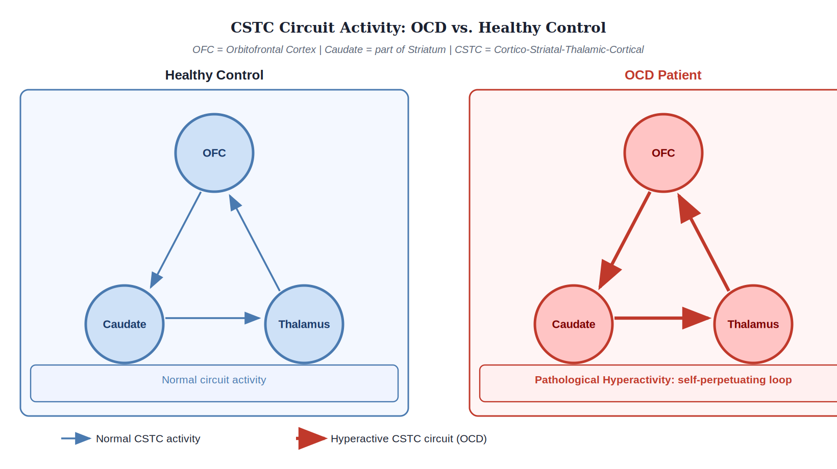
The figure shows a simplified model of brain activity in an individual with Obsessive-Compulsive Disorder (OCD) compared to a healthy control while both are exposed to a symptom-provoking stimulus. Based on the model shown in the figure, which statement best describes the neural mechanism underlying the patient's symptoms?
- The patient's amygdala is sending excessive fear signals to the cortex, a pattern typical of specific phobia.
- The patient has deficient top-down control from the cingulate cortex, leading to general disinhibition of the limbic system.
- The patient shows widespread, non-specific hyperactivity, indicating a general state of anxiety rather than a specific circuit dysfunction.
- The patient is experiencing a self-perpetuating loop of hyperactivity in the cortico-striatal-thalamic-cortical circuit. (correct answer)
Explanation: The correct answer is D. The figure depicts hyperactivity specifically in the orbitofrontal cortex (OFC), caudate nucleus (striatum), and thalamus, which form the core of the cortico-striatal-thalamic-cortical (CSTC) circuit. This circuit is the key neural substrate implicated in OCD. The model shows this loop as pathologically overactive, representing the intrusive obsessions and compulsive urges that fail to terminate, which is the leading neurobiological explanation for OCD symptoms. (A) is incorrect because the amygdala is not highlighted, and the pattern is specific to OCD, not phobias. (B) is too general and does not account for the specific loop shown. (C) is incorrect because the figure shows highly specific and localized hyperactivity, not a widespread pattern.
Question 2
A combat veteran with Post-Traumatic Stress Disorder (PTSD) experiences intrusive, emotionally-charged flashbacks that are often fragmented and lack clear spatial-temporal context. This specific symptomatology is best explained by the neurobiological interaction between:
- a hyperactive amygdala strengthening emotional memory consolidation and a dysfunctional hippocampus failing to properly contextualize the memory. (correct answer)
- a hypoactive prefrontal cortex failing to inhibit motor impulses and an overactive cerebellum creating procedural memory errors.
- a dysfunctional thalamus failing to gate sensory information and a hyperactive occipital lobe creating vivid visual distortions.
- a dysregulated hypothalamus disrupting the stress response and an atrophied basal ganglia impairing habit formation.
Explanation: The correct answer is A. The core features of PTSD flashbacks involve both intense emotion and disorganized memory. The amygdala is central to processing fear and attaching emotional significance to memories, and its hyperactivity in PTSD leads to the intense emotional charge. The hippocampus is crucial for forming coherent, contextualized episodic memories. Dysfunction or reduced volume in the hippocampus, often seen in PTSD, impairs the ability to place the traumatic memory in a specific time and place, leading to its fragmented and intrusive nature. (B), (C), and (D) list brain regions involved in other functions that are less central to this specific memory-related symptom of PTSD.
Question 3
A patient beginning treatment with a selective serotonin reuptake inhibitor (SSRI) for major depression reports feeling no improvement after one week, even though the drug has already increased synaptic serotonin levels. The most widely accepted neurological explanation for the typical multi-week delay in the therapeutic effects of SSRIs is that they depend on:
- the slow process of the drug crossing the blood-brain barrier and reaching a therapeutic concentration in the cerebrospinal fluid.
- the gradual desensitization of presynaptic serotonin autoreceptors, which initially counteract the increase in serotonin release.
- the time required for the patient's brain to metabolize the drug into its active compounds, which varies based on liver function.
- the initiation of slower, downstream neuroadaptive changes, such as altered gene expression and increased hippocampal neurogenesis. (correct answer)
Explanation: The correct answer is D. While SSRIs increase synaptic serotonin levels quickly, their antidepressant effects are not immediate. The leading hypothesis for this delay is that the sustained increase in serotonin initiates slower, downstream processes. These include changes in the expression of genes related to neuronal growth and survival (like BDNF) and promoting the growth of new neurons (neurogenesis), particularly in the hippocampus, a brain region implicated in depression. (A) and (C) are incorrect as drug concentration reaches a steady state much faster, typically within a week. (B) is a real phenomenon that occurs, but it is considered an early-stage adaptation and not the full explanation for the weeks-long therapeutic delay, which is better explained by the more profound neuroplastic changes.
Question 4
After surviving an accident where a metal rod passed through his skull, a man's memory and general intelligence remain unimpaired, but his personality changes dramatically. He becomes profane, impulsive, and unable to make rational decisions about his social life or finances. This specific profile of deficits strongly suggests the injury caused significant damage to his:
- bilateral temporal lobes, including the hippocampus.
- parietal association cortex.
- ventromedial and orbitofrontal prefrontal cortex. (correct answer)
- primary motor and somatosensory cortices.
Explanation: The correct answer is C. This case is a classic example based on the historical account of Phineas Gage. The ventromedial and orbitofrontal regions of the prefrontal cortex are crucial for integrating emotion into decision-making, social conduct, and risk assessment. Damage to this area leaves cognitive functions like memory and intelligence intact but severely impairs judgment, impulse control, and the ability to plan for the future, leading to the personality changes described. (A) would cause severe memory problems (amnesia). (B) would lead to spatial neglect or other deficits in spatial processing. (D) would cause paralysis or loss of sensation in parts of the body.
Question 5
Tourette Syndrome is a neurodevelopmental disorder characterized by involuntary motor and vocal tics. The prevailing neurobiological model suggests that these tics arise from dysfunction within the cortico-striatal-thalamo-cortical loops. Specifically, this dysfunction is thought to result in a:
- failure of the basal ganglia to adequately inhibit competing, unwanted motor programs. (correct answer)
- hypersensitivity of acetylcholine receptors at the neuromuscular junction.
- developmental lesion in the cerebellum that disrupts the smoothness of motor sequences.
- chronic overactivation of the primary motor cortex due to faulty sensory gating in the thalamus.
Explanation: The correct answer is A. The basal ganglia, a key part of the CSTC loops, normally acts as a gatekeeper, inhibiting unwanted movements while allowing desired ones to proceed. In Tourette Syndrome, this inhibitory function is thought to be impaired. This leads to the spontaneous release of fragments of motor programs, which manifest as tics. Thus, tics are not caused by a desire to move, but by a failure of the brain's braking system. (B) describes a peripheral issue, not a central one. (C) would lead to ataxia (uncoordinated movement), not tics. (D) is an oversimplification that misses the critical role of the basal ganglia's inhibitory function.
Question 6
Repetitive transcranial magnetic stimulation (rTMS) is an FDA-approved treatment for medication-resistant major depression. The standard protocol involves using a magnetic coil to induce electrical currents in the brain. The therapeutic effect is believed to result from stimulating neurons in the:
- hippocampus, because it is a region known to exhibit reduced volume in depressed patients.
- amygdala, in order to directly suppress the brain's fear and anxiety response center.
- left dorsolateral prefrontal cortex, a region often showing hypoactivity in depression. (correct answer)
- primary motor cortex, which is functionally connected to emotional regulation circuits.
Explanation: The correct answer is C. The most common and well-studied protocol for rTMS for depression involves applying high-frequency (excitatory) stimulation to the left dorsolateral prefrontal cortex (DLPFC). This specific area is targeted because neuroimaging studies have consistently shown it to be underactive in individuals with depression. The goal of the treatment is to normalize activity in this region and its connected networks, thereby alleviating depressive symptoms. (A) and (B) are incorrect because these structures are too deep in the brain to be directly and non-invasively stimulated by TMS. (D) is incorrect because while the motor cortex is used to calibrate the stimulation strength, it is not the therapeutic target for depression.
Question 7
A patient with panic disorder is prescribed a benzodiazepine for short-term relief. The drug's anxiolytic effect is achieved by positively modulating the brain's main inhibitory neurotransmitter system. At the synaptic level, this drug facilitates the binding of GABA to its receptor, which directly results in an:
- increased influx of Cl⁻ ions, causing neuronal hyperpolarization. (correct answer)
- increased efflux of K⁺ ions, causing neuronal hyperpolarization.
- decreased influx of Na⁺ ions, preventing neuronal depolarization.
- decreased efflux of Ca²⁺ ions, preventing neurotransmitter release.
Explanation: The correct answer is A. Benzodiazepines are positive allosteric modulators of the GABA-A receptor. When GABA binds to this receptor, it opens a channel that allows negatively charged chloride (Cl⁻) ions to flow into the neuron. Benzodiazepines enhance this effect, causing an even greater influx of Cl⁻. This makes the neuron's membrane potential more negative (hyperpolarization), moving it further from the threshold for firing an action potential, thus producing a widespread inhibitory effect. (B) describes the action of potassium channels, not the GABA-A receptor. (C) and (D) describe different ionic mechanisms not directly related to the action of benzodiazepines on GABA-A receptors.
Question 8
A patient treated for several years with a first-generation (typical) antipsychotic for schizophrenia develops tardive dyskinesia, characterized by involuntary facial grimacing and tongue protrusions. This serious motor side effect is a direct consequence of the medication's chronic blockade of D2 dopamine receptors in the:
- mesolimbic pathway, leading to anhedonia.
- mesocortical pathway, worsening negative symptoms.
- nigrostriatal pathway, causing receptor supersensitivity. (correct answer)
- tuberoinfundibular pathway, leading to hyperprolactinemia.
Explanation: The correct answer is C. First-generation antipsychotics are not selective and block D2 dopamine receptors throughout the brain. While their intended therapeutic effect comes from blocking these receptors in the mesolimbic pathway (to reduce positive symptoms), their blockade in the nigrostriatal pathway disrupts normal motor function. Over time, the brain tries to compensate for this chronic blockade by increasing the number and sensitivity of D2 receptors in this pathway. This supersensitivity is thought to cause the hyperkinetic, involuntary movements of tardive dyskinesia. (A) and (B) describe effects in other pathways. (D) describes another common side effect of these drugs, but it is unrelated to the motor symptoms of tardive dyskinesia.
Question 9
Beyond the well-known dopamine hypothesis, a compelling alternative model suggests that the negative symptoms and cognitive deficits of schizophrenia may stem from the hypofunction of a different neurotransmitter system. This model is supported by the psychotomimetic effects of drugs like phencyclidine (PCP), which act as antagonists for:
- opioid receptors.
- NMDA-type glutamate receptors. (correct answer)
- muscarinic acetylcholine receptors.
- alpha-adrenergic norepinephrine receptors.
Explanation: The correct answer is B. This question refers to the glutamate hypothesis of schizophrenia. Glutamate is the brain's primary excitatory neurotransmitter, and the NMDA receptor is a key type of glutamate receptor. The fact that NMDA receptor antagonists like PCP and ketamine can produce symptoms that closely resemble the negative and cognitive aspects of schizophrenia provides strong evidence for this hypothesis. It suggests that a primary deficit in glutamate signaling (hypofunction) may underlie some of the core features of the disorder that are poorly explained by the dopamine hypothesis alone. The other options list receptor systems not central to this particular model of schizophrenia.
Question 10
A clinician observes that a patient with schizophrenia exhibits significant negative symptoms, such as avolition and alogia, with minimal positive symptoms like hallucinations. This specific symptom profile is most strongly associated with dysfunction in which specific neural pathway?
- Hyperactivity in the mesolimbic dopamine pathway, which projects from the VTA to the nucleus accumbens.
- Hypoactivity in the mesocortical dopamine pathway, which projects from the VTA to the prefrontal cortex. (correct answer)
- Degeneration of the nigrostriatal pathway, which projects from the substantia nigra to the striatum.
- Dysregulation of the tuberoinfundibular pathway, which projects from the hypothalamus to the pituitary gland.
Explanation: The correct answer is B. The dopamine hypothesis of schizophrenia has been refined to link different pathways to different symptoms. Negative symptoms (e.g., lack of motivation, poverty of speech) and cognitive deficits are thought to be related to a deficit of dopamine, or hypoactivity, in the mesocortical pathway that projects to the prefrontal cortex. (A) is incorrect because hyperactivity in the mesolimbic pathway is associated with the positive symptoms of schizophrenia, such as hallucinations and delusions. This is a common point of confusion. (C) is incorrect because degeneration of the nigrostriatal pathway is the primary cause of Parkinson's disease. (D) is incorrect because the tuberoinfundibular pathway is involved in regulating prolactin, and its disruption by antipsychotic medications can cause side effects but is not linked to the primary symptoms of schizophrenia.
Question 11
Neuroimaging studies of bipolar disorder often reveal a pattern of brain abnormalities that helps explain the core symptom of emotional dysregulation. Which of the following findings best represents the typical push-pull imbalance between neural systems thought to underlie this symptom?
- Enlarged ventricles, suggesting widespread neuronal loss primarily in subcortical regions.
- Reduced prefrontal cortical volume and activity, coupled with amygdala hyperactivity. (correct answer)
- Degeneration of dopamine neurons in the substantia nigra, leading to motor system instability.
- Formation of amyloid plaques in the hippocampus, disrupting memory and emotional processing.
Explanation: The correct answer is B. A key neurobiological model of bipolar disorder posits a functional imbalance between emotion-generating brain regions (like the amygdala) and emotion-regulating regions (like the prefrontal cortex). Studies consistently find that individuals with bipolar disorder show hyperactivity in the amygdala in response to emotional stimuli, along with reduced gray matter volume and diminished activity in parts of the prefrontal cortex responsible for top-down emotional control. This combination is thought to underlie the mood instability and impulsivity seen in both manic and depressive states. (A) is more characteristic of schizophrenia. (C) is the hallmark of Parkinson's disease. (D) is the hallmark of Alzheimer's disease.
Question 12
A researcher is studying the neurobiology of cocaine addiction. While the drug affects dopamine levels throughout the mesolimbic pathway, its intensely reinforcing and 'high'-producing effects are most directly mediated by a rapid and substantial increase of dopamine concentration specifically within the:
- ventral tegmental area (VTA), where dopamine neurons originate.
- nucleus accumbens, which is the primary target of VTA projections. (correct answer)
- prefrontal cortex, which modulates craving and decision-making.
- hippocampus, which forms contextual memories of drug use.
Explanation: The correct answer is B. The nucleus accumbens is a critical component of the brain's reward circuitry and the primary site where the rewarding effects of most drugs of abuse are mediated. Dopamine neurons originating in the VTA project to the nucleus accumbens. Drugs like cocaine block dopamine reuptake, causing a surge of dopamine specifically in the synapses of the nucleus accumbens, which is experienced as euphoria and reinforcement. (A) is incorrect because the VTA is the source of the dopamine neurons, not the primary site of the rewarding effect. (C) and (D) are involved in the broader context of addiction (craving, memory), but the acute rewarding effect is centered in the nucleus accumbens.
Question 13
Cholinesterase inhibitors are a class of drugs commonly prescribed to manage the cognitive symptoms of early-to-moderate Alzheimer's disease. These drugs work by increasing the availability of acetylcholine in the synapse. This treatment temporarily compensates for the degeneration of cholinergic neurons that originate primarily in which subcortical region?
- Ventral tegmental area
- Raphe nuclei
- Basal forebrain (correct answer)
- Locus coeruleus
Explanation: The correct answer is C. One of the earliest neurochemical changes in Alzheimer's disease is the loss of acetylcholine-producing neurons. The primary source of acetylcholine for the entire cerebral cortex is a collection of structures in the basal forebrain, including the nucleus basalis of Meynert. Cholinesterase inhibitors work by blocking the enzyme that breaks down acetylcholine, thus increasing its levels and compensating for this neuronal loss. (A) is the primary source of dopamine for the reward pathway. (B) is the primary source of serotonin. (D) is the primary source of norepinephrine.
Question 14
Cholinesterase inhibitors are a class of drugs commonly prescribed to manage the cognitive symptoms of early-to-moderate Alzheimer's disease. These drugs work by increasing the availability of acetylcholine in the synapse. This treatment temporarily compensates for the degeneration of cholinergic neurons that originate primarily in which subcortical region?
- Ventral tegmental area
- Raphe nuclei
- Basal forebrain (correct answer)
- Locus coeruleus
Explanation: The correct answer is C. One of the earliest neurochemical changes in Alzheimer's disease is the loss of acetylcholine-producing neurons. The primary source of acetylcholine for the entire cerebral cortex is a collection of structures in the basal forebrain, including the nucleus basalis of Meynert. Cholinesterase inhibitors work by blocking the enzyme that breaks down acetylcholine, thus increasing its levels and compensating for this neuronal loss. (A) is the primary source of dopamine for the reward pathway. (B) is the primary source of serotonin. (D) is the primary source of norepinephrine.
Question 15
The 'stress-diathesis' model of depression posits that chronic stress can be a significant contributing factor to the disorder. Prolonged activation of the hypothalamic-pituitary-adrenal (HPA) axis and the resulting high levels of cortisol are believed to be neurotoxic, contributing to depressive symptoms by causing which of the following cellular changes?
- Increased rate of myelination in cortical neurons, leading to inefficient signal processing.
- Upregulation of dopamine receptors in the nucleus accumbens, leading to anhedonia.
- Atrophy of neuronal dendrites and inhibition of neurogenesis, particularly in the hippocampus. (correct answer)
- Massive apoptosis of GABAergic interneurons in the amygdala, leading to hyperexcitability.
Explanation: The correct answer is C. The hippocampus, which plays a key role in both memory and the regulation of the stress response, is particularly vulnerable to the effects of chronic stress and high cortisol levels. Research shows that prolonged stress can cause the dendrites of hippocampal neurons to shrink (atrophy) and can suppress the birth of new neurons (neurogenesis) in this region. This structural impairment is thought to contribute to the cognitive and mood symptoms of depression. (A) is incorrect; stress does not increase myelination. (B) describes a change more related to addiction and is the opposite of what would cause anhedonia. (D) is an extreme outcome not typically associated with chronic stress in depression models.
Question 16
During a panic attack, an individual experiences an abrupt and intense surge of autonomic arousal, including tachycardia and shortness of breath. While the amygdala is critical for processing the 'fear' component, this rapid, widespread physiological alarm is triggered primarily by a burst of norepinephrine released from the:
- substantia nigra.
- locus coeruleus. (correct answer)
- raphe nuclei.
- reticular formation.
Explanation: The correct answer is B. The locus coeruleus, located in the pons region of the brainstem, is the brain's principal site for synthesizing norepinephrine. It has extensive projections throughout the brain and spinal cord and plays a primary role in mediating the physiological responses to stress and panic (the 'fight-or-flight' response). A sudden, massive burst of activity in the locus coeruleus is thought to initiate the powerful physical symptoms of a panic attack. (A) is a source of dopamine for the motor system. (C) is a source of serotonin. (D) is involved in general arousal and consciousness, but the locus coeruleus is the more specific origin of the sympathetic nervous system alarm signal.
Question 17
Individuals with anorexia nervosa often exhibit a profound disconnect between their internal state and their perception of that state, such as not 'feeling' hungry despite physiological starvation. This impairment in interoception—the sensing of the body's internal condition—is most strongly associated with altered structure and function in which cortical region?
- The fusiform gyrus
- The hypothalamus
- The cerebellum
- The insular cortex (correct answer)
Explanation: The correct answer is D. The insular cortex, or insula, is a key brain region for interoception. It integrates signals from within the body (e.g., from the heart, lungs, gut) to create a conscious sense of our internal physiological state, including hunger, pain, and emotional feelings. Altered activity in the insula is a consistent finding in studies of anorexia nervosa and is thought to underlie the distorted awareness of hunger and body state that characterizes the disorder. (A) is involved in object and face recognition. (B), while critical for the homeostatic regulation of hunger, is not the primary site for the conscious perception of that hunger. (C) is primarily involved in motor control.
Question 18
The neurological mechanism of exposure therapy for specific phobias involves fear extinction learning. Successful therapy is characterized by a re-wiring of brain circuits such that the fear response is suppressed. This is primarily accomplished through a strengthening of inhibitory, top-down control from the over the activity of the .
- hippocampus; thalamus
- amygdala; brainstem
- ventromedial prefrontal cortex (vmPFC); amygdala (correct answer)
- sensory cortex; ventromedial prefrontal cortex (vmPFC)
Explanation: The correct answer is C. Fear extinction is not about erasing the original fear memory but about learning a new, competing memory that the feared stimulus is safe. The ventromedial prefrontal cortex (vmPFC) is critical for this process. During successful exposure therapy, the vmPFC becomes more active and exerts inhibitory control over the amygdala, which is the brain's fear center. This top-down regulation from the vmPFC effectively dampens the amygdala's fear output, leading to a reduction in the phobic response. The other options describe incorrect pathways or reverse the direction of control for fear extinction.
Question 19
Tourette Syndrome is a neurodevelopmental disorder characterized by involuntary motor and vocal tics. The prevailing neurobiological model suggests that these tics arise from dysfunction within the cortico-striatal-thalamo-cortical loops. Specifically, this dysfunction is thought to result in a:
- failure of the basal ganglia to adequately inhibit competing, unwanted motor programs. (correct answer)
- hypersensitivity of acetylcholine receptors at the neuromuscular junction.
- developmental lesion in the cerebellum that disrupts the smoothness of motor sequences.
- chronic overactivation of the primary motor cortex due to faulty sensory gating in the thalamus.
Explanation: The correct answer is A. The basal ganglia, a key part of the CSTC loops, normally acts as a gatekeeper, inhibiting unwanted movements while allowing desired ones to proceed. In Tourette Syndrome, this inhibitory function is thought to be impaired. This leads to the spontaneous release of fragments of motor programs, which manifest as tics. Thus, tics are not caused by a desire to move, but by a failure of the brain's braking system. (B) describes a peripheral issue, not a central one. (C) would lead to ataxia (uncoordinated movement), not tics. (D) is an oversimplification that misses the critical role of the basal ganglia's inhibitory function.
Question 20
A patient with advanced Parkinson's disease, whose motor tremors are no longer controlled by L-DOPA medication, undergoes deep brain stimulation (DBS). To be most effective at alleviating these specific motor symptoms, the electrodes should be implanted to directly modulate activity within which neural circuit?
- The amygdalo-hippocampal circuit, to reduce the anxiety and cognitive impairment sometimes associated with the disease.
- The mesolimbic dopamine pathway, to compensate for the general loss of dopamine throughout the brain.
- The cerebellar-pontine pathway, to improve the timing and coordination of voluntary movements.
- The cortico-basal ganglia-thalamo-cortical loop, specifically targeting the subthalamic nucleus or globus pallidus. (correct answer)
Explanation: The correct answer is D. The motor symptoms of Parkinson's disease are caused by the degeneration of dopamine-producing neurons in the substantia nigra, which leads to dysregulation of the cortico-basal ganglia-thalamo-cortical motor circuit. Deep brain stimulation (DBS) targets key nodes within this circuit, such as the subthalamic nucleus or globus pallidus, to restore more normal patterns of activity and alleviate tremors and rigidity. (A) is incorrect because the amygdala and hippocampus are involved in emotion and memory, not the primary motor symptoms of Parkinson's. (B) is incorrect because while Parkinson's involves dopamine loss, DBS is a circuit-based intervention, and the mesolimbic pathway is more associated with reward and addiction. (C) is incorrect because the cerebellum is involved in motor coordination, but the primary pathology of Parkinson's and the target of DBS is the basal ganglia circuit.