All questions
Question 1
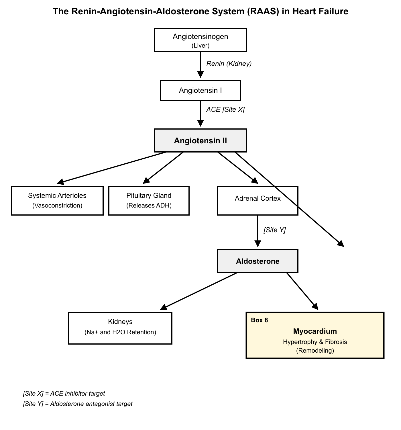
Based on the provided diagram of the RAAS pathway, a patient with HFrEF is treated with a medication that acts at [Site Y]. In addition to blocking the effects on preload, this therapy has been shown to directly attenuate the pathological process occurring at Box 8. Which statement best explains this secondary benefit?
- The drug provides negative feedback to the kidney, suppressing renin release and thereby reducing the formation of all downstream products including Angiotensin II.
- By acting at [Site Y], the drug prevents the conversion of Angiotensin I to Angiotensin II, which is the primary stimulus for the process in Box 8.
- The drug, an aldosterone antagonist, directly blocks mineralocorticoid receptors on cardiac fibroblasts and myocytes, reducing collagen synthesis and hypertrophy. (correct answer)
- The medication enhances the degradation of Angiotensin II, preventing its vasoconstrictive effects and reducing cardiac afterload, which lessens the stimulus for remodeling.
Explanation: Correct: Site Y is downstream of aldosterone, indicating a blockade of aldosterone's effects. Box 8 represents myocardial remodeling (hypertrophy and fibrosis). An aldosterone antagonist (like spironolactone or eplerenone) acts at Site Y. Its benefit extends beyond its diuretic effect (reducing preload). Aldosterone has direct effects on the heart by binding to mineralocorticoid receptors on cardiomyocytes and fibroblasts, promoting inflammation, fibrosis, and hypertrophy. Blocking these receptors directly attenuates this adverse remodeling.
A: Blocking aldosterone's effects can lead to a compensatory increase in renin release, not suppression.
B: This describes the action of an ACE inhibitor, which would act at [Site X] on the diagram.
D: This describes an incorrect mechanism of action; aldosterone antagonists do not enhance Angiotensin II degradation.
Question 2
A patient with heart failure due to a previous myocardial infarction develops a dilated left ventricle with a thinned wall (eccentric hypertrophy). A second patient with heart failure from long-standing hypertension develops a thickened left ventricular wall with a reduced chamber diameter (concentric hypertrophy). Which statement best describes the initial hemodynamic stimuli that drive these distinct remodeling patterns?
- Eccentric hypertrophy is primarily a response to chronic pressure overload, while concentric hypertrophy is a response to chronic volume overload.
- Both remodeling patterns are driven primarily by direct catecholamine-induced myocyte apoptosis, with the pattern determined by the location of cell death.
- Concentric hypertrophy is an adaptive response to increased wall stress from pressure overload, while eccentric hypertrophy results from volume overload and neurohormonal activation following loss of contractile tissue. (correct answer)
- Eccentric hypertrophy is caused by aldosterone-mediated fibrosis, while concentric hypertrophy is caused by angiotensin II-mediated myocyte growth.
Explanation: Correct: These two patterns of remodeling are driven by different initial insults. Concentric hypertrophy (thickened wall) is a classic response to chronic pressure overload (like hypertension), where sarcomeres are added in parallel to reduce wall stress. Eccentric hypertrophy (dilated chamber) is a response to volume overload or loss of myocardium (like in an MI), where new sarcomeres are added in series to accommodate the increased volume, leading to chamber dilation. Neurohormonal activation contributes significantly to both processes long-term, but the initial stimulus is key.
A: This reverses the stimuli; pressure overload leads to concentric, and volume overload to eccentric.
B: While apoptosis occurs, it doesn't primarily determine the geometric pattern of hypertrophy.
D: This is an oversimplification; both aldosterone and Ang II contribute to fibrosis and hypertrophy in both patterns of remodeling.
Question 3
Cardiac remodeling in response to pressure overload, such as in aortic stenosis, involves significant changes in the extracellular matrix. Which statement accurately describes the roles of angiotensin II (Ang II) and norepinephrine (NE) in this specific remodeling process?
- NE is the primary stimulus for fibroblast proliferation and collagen synthesis, while Ang II primarily mediates myocyte hypertrophy.
- NE reduces cardiac fibrosis by inhibiting transforming growth factor-beta (TGF-β), while Ang II promotes fibrosis via the same pathway.
- Ang II mediates a switch in myocyte gene expression to a fetal pattern, while NE is primarily responsible for myocyte apoptosis.
- Ang II and NE both act as potent growth factors for cardiomyocytes and fibroblasts, stimulating myocyte hypertrophy and interstitial fibrosis. (correct answer)
Explanation: When you encounter questions about cardiac remodeling in pressure overload conditions like aortic stenosis, focus on understanding how neurohormonal activation drives both cardiomyocyte and fibroblast responses. The heart responds to increased workload by activating the sympathetic nervous system and renin-angiotensin system, releasing norepinephrine and angiotensin II as key mediators.
Both angiotensin II and norepinephrine function as potent growth factors in cardiac remodeling. They stimulate cardiomyocyte hypertrophy through activation of protein kinase pathways and increased protein synthesis. Simultaneously, both hormones promote fibroblast proliferation and collagen synthesis, leading to interstitial fibrosis. This dual action on both cell types makes answer D correct – both mediators contribute significantly to myocyte hypertrophy and fibrosis formation.
Answer A incorrectly suggests these mediators have distinct, non-overlapping roles, when in reality both contribute to fibroblast activation and myocyte growth. Answer B contains a major error – norepinephrine does not reduce fibrosis or inhibit TGF-β; it actually promotes fibrotic changes. Answer C mischaracterizes their primary effects. While angiotensin II does influence fetal gene expression patterns, this isn't its primary distinguishing role, and norepinephrine's main effect isn't apoptosis but rather hypertrophy and fibrosis promotion.
Remember that in cardiac pathophysiology, neurohormonal mediators typically have overlapping, synergistic effects rather than completely distinct roles. Both sympathetic activation (norepinephrine) and RAAS activation (angiotensin II) work together to drive the maladaptive remodeling process in heart failure.
Question 4
A 70-year-old female with HFrEF (EF 25%) is admitted with acute decompensation, presenting with severe dyspnea and hypotension (BP 85/60 mmHg). She is on an ACE inhibitor, a beta-blocker, and a diuretic. In this acute, low-output state, which compensatory mechanism is most responsible for maintaining perfusion to her brain and heart at the expense of other organs?
- A profound surge in B-type natriuretic peptide (BNP) causing selective vasodilation of coronary and cerebral arteries.
- Aldosterone-mediated volume retention, which rapidly increases circulating volume to raise systemic blood pressure.
- Activation of the parasympathetic nervous system, causing bradycardia to improve diastolic filling and coronary perfusion.
- Intense sympathetic nervous system (SNS) activation leading to potent vasoconstriction in the splanchnic, renal, and cutaneous circulations. (correct answer)
Explanation: When you encounter a heart failure question describing acute decompensation with hypotension, focus on the body's immediate compensatory responses. In this low-output state, the cardiovascular system must rapidly redistribute blood flow to protect vital organs.
The correct answer is D because intense sympathetic activation is the primary acute compensatory mechanism in cardiogenic shock. When cardiac output drops severely (EF 25% with hypotension), baroreceptors detect the pressure drop and trigger massive SNS activation. This causes potent vasoconstriction in non-essential vascular beds—specifically the splanchnic (gut), renal, and cutaneous circulations—while relatively sparing coronary and cerebral vessels. This redistribution maintains perfusion pressure to the brain and heart at the expense of other organs, explaining why patients develop cool, clammy skin and may develop acute kidney injury.
Option A is incorrect because while BNP does rise in heart failure, it doesn't cause selective vasodilation of vital organs—it actually causes generalized vasodilation and diuresis, which would worsen hypotension in this acute setting.
Option B is wrong because aldosterone-mediated volume retention is a chronic compensatory mechanism that takes hours to days to affect blood pressure, not minutes to hours needed in acute decompensation.
Option C is incorrect because parasympathetic activation would be counterproductive here—bradycardia would further reduce cardiac output when the heart is already failing.
Remember: In acute heart failure with hypotension, think "sympathetic storm"—the body's immediate response is always intense SNS activation to maintain vital organ perfusion through selective vasoconstriction.
Question 5
A patient with chronic heart failure with reduced ejection fraction (HFrEF) exhibits decreased responsiveness to an intravenous dobutamine infusion. This blunted response is primarily a consequence of long-term compensatory sympathetic nervous system (SNS) activation. Which cellular mechanism is the most direct cause of this phenomenon?
- Depletion of intracellular calcium stores from the sarcoplasmic reticulum, impairing the final step of excitation-contraction coupling.
- Chronic elevation of circulating catecholamines leading to downregulation of β1-adrenergic receptors and increased G-protein-coupled receptor kinase (GRK) activity. (correct answer)
- Competitive inhibition of dobutamine binding by elevated levels of endogenous B-type natriuretic peptide (BNP) at the receptor site.
- Increased myocardial expression of phosphodiesterase-3, which accelerates the degradation of cyclic GMP (cGMP) and shortens the inotropic response.
Explanation: Correct: In chronic HF, persistent SNS activation leads to high levels of norepinephrine, which causes β1-adrenergic receptors to be phosphorylated by GRKs. This marks them for internalization (downregulation) and uncouples them from their G-protein (desensitization), reducing the heart's responsiveness to both endogenous catecholamines and exogenous agonists like dobutamine.
A: While calcium handling is abnormal in HF, the primary reason for blunted response to a β-agonist is receptor-level changes, not depletion of the stores themselves.
C: BNP is a counter-regulatory hormone and does not competitively inhibit β-adrenergic receptors.
D: Phosphodiesterases in the heart primarily degrade cyclic AMP (cAMP), not cGMP, to terminate the β-adrenergic signal. Increased activity would blunt the response, but downregulation of the receptor itself is the more fundamental, chronic adaptation.
Question 6
Patients with advanced heart failure have an increased risk of ventricular arrhythmias. While multiple factors are involved, chronic SNS activation is a major contributor. Which mechanism best explains how sustained high levels of catecholamines promote arrhythmogenesis in the failing myocardium?
- By increasing parasympathetic tone, which leads to unpredictable changes in sinoatrial node firing rates and atrioventricular conduction.
- By causing hyperkalemia through direct effects on renal tubules, which lowers the resting membrane potential of cardiomyocytes and increases their excitability.
- By promoting hypokalemia and increasing late sodium current and calcium influx into myocytes, which can lead to early and delayed afterdepolarizations. (correct answer)
- By stimulating myocardial fibrosis, which insulates electrical conduction and prevents the formation of re-entrant circuits.
Explanation: Correct: Chronic sympathetic stimulation has several pro-arrhythmic effects. It can cause hypokalemia (by stimulating beta-2 receptors that shift potassium into cells). More directly, it increases intracellular calcium by enhancing L-type calcium channel current and sarcoplasmic reticulum calcium release. This calcium overload can trigger delayed afterdepolarizations (DADs), which are abnormal depolarizations that can initiate ventricular tachycardia. Catecholamines can also prolong the action potential duration, predisposing to early afterdepolarizations (EADs).
A: SNS activation leads to parasympathetic withdrawal, not increased tone.
B: SNS activation tends to cause hypokalemia, not hyperkalemia.
D: Myocardial fibrosis creates areas of slow conduction and conduction block, which is a substrate that promotes, not prevents, the formation of re-entrant circuits for arrhythmias.
Question 7
Cardiomyocyte apoptosis is a critical feature of adverse cardiac remodeling in heart failure. Sustained neurohormonal stress from both the SNS and RAAS activates intracellular pathways that lead to this programmed cell death. Which molecular mechanism is a key link between these compensatory systems and apoptosis?
- Norepinephrine and angiotensin II both suppress the activity of pro-apoptotic proteins like Bax and Bad.
- Natriuretic peptides, in their attempt to be cardioprotective, inadvertently activate caspase-3, leading to widespread cell death.
- Aldosterone directly activates death receptors like Fas on the cardiomyocyte surface, initiating the extrinsic apoptosis pathway.
- Activation of β1-adrenergic and AT1 receptors leads to sustained increases in intracellular Ca²⁺ and reactive oxygen species (ROS), which trigger mitochondrial-mediated apoptosis. (correct answer)
Explanation: When you encounter questions about heart failure pathophysiology, focus on how compensatory mechanisms eventually become harmful through sustained activation. The key insight is understanding how the sympathetic nervous system (SNS) and renin-angiotensin-aldosterone system (RAAS) initially help maintain cardiac output but ultimately damage the heart through chronic overstimulation.
The correct mechanism linking neurohormonal stress to cardiomyocyte death involves β1-adrenergic and AT1 receptor activation leading to calcium overload and oxidative stress. When norepinephrine binds β1-receptors and angiotensin II binds AT1 receptors, both pathways increase intracellular calcium through different mechanisms—β1 via cAMP and AT1 via IP3/DAG signaling. This sustained calcium elevation, combined with increased reactive oxygen species production, damages mitochondria and triggers the intrinsic apoptosis pathway. The mitochondria release cytochrome c, activating caspases and ultimately causing programmed cell death. This makes option D correct.
Option A is backwards—norepinephrine and angiotensin II actually promote apoptosis by activating pro-apoptotic proteins, not suppressing them. Option B incorrectly implicates natriuretic peptides, which are genuinely cardioprotective and don't activate caspase-3. Option C mischaracterizes aldosterone's mechanism; while aldosterone does contribute to cardiac damage, it doesn't directly activate Fas death receptors on cardiomyocytes.
Remember that in heart failure pathophysiology, compensatory mechanisms follow a predictable pattern: short-term benefit followed by long-term harm through receptor overstimulation, calcium dysregulation, and oxidative stress leading to cell death.
Question 8
A patient with heart failure is treated with an ACE inhibitor. While this effectively lowers angiotensin II levels initially, after several months of therapy, aldosterone levels are found to have returned to baseline despite continued ACE inhibition. This demonstrates a limited long-term effect of monotherapy. Which compensatory change most likely accounts for this 'aldosterone escape'?
- Increased production of angiotensin II through non-ACE pathways, such as chymase in the heart, which continues to stimulate the adrenal cortex. (correct answer)
- Downregulation of ACE receptors in the lung, leading to decreased drug efficacy and a rebound in angiotensin II production.
- Direct stimulation of aldosterone release by elevated B-type natriuretic peptide (BNP) levels, which bypasses the need for angiotensin II.
- A drug-induced increase in renal potassium retention, which directly suppresses aldosterone synthase activity in the adrenal gland.
Explanation: Correct: ACE is the primary, but not the only, enzyme that can convert angiotensin I to angiotensin II. Other enzymes, such as chymase, are present in tissues like the heart and can perform this conversion. In the setting of chronic ACE inhibition, these alternative pathways can become more active, leading to a gradual rise in local and systemic angiotensin II levels, which then stimulate the adrenal gland to produce aldosterone. This is a key reason why direct aldosterone antagonists are beneficial even in patients on ACE inhibitors.
B: ACE is an enzyme, not a receptor, and it's not the primary mechanism.
C: BNP inhibits, rather than stimulates, aldosterone release.
D: Hyperkalemia (potassium retention) inhibits, not stimulates, aldosterone release. This is a distractor that correctly identifies a side effect of ACE inhibitors but misapplies its effect on aldosterone.
Question 9
A 68-year-old male with decompensated systolic heart failure presents with 3+ pitting edema in his lower extremities, ascites, and cool, clammy skin. Which combination of compensatory mechanisms best explains this specific constellation of signs?
- Activation of natriuretic peptides causing vasodilation (cool skin) and RAAS activation causing sodium retention (edema).
- Sympathetic nervous system activation causing peripheral vasoconstriction (cool skin) and RAAS activation causing sodium and water retention (edema). (correct answer)
- Release of ADH causing free water retention (edema) and downregulation of β-receptors causing reduced cardiac output (cool skin).
- Increased parasympathetic tone causing bradycardia (cool skin) and aldosterone escape causing volume overload (edema).
Explanation: Correct: This question requires integrating the effects of the two major compensatory systems. The edema and ascites are classic signs of volume overload caused by the renin-angiotensin-aldosterone system (RAAS), which promotes renal sodium and water retention. The cool, clammy skin is a sign of peripheral vasoconstriction, a mechanism mediated by the sympathetic nervous system (SNS) to shunt blood from the periphery to vital organs in a low cardiac output state.
A: Natriuretic peptides cause vasodilation, but this would lead to warm, not cool, skin.
C: β-receptor downregulation is a chronic change and doesn't directly cause the acute sign of cool skin; acute SNS-mediated vasoconstriction does.
D: Heart failure is characterized by decreased, not increased, parasympathetic tone.
Question 10
A patient with HFrEF is transitioned from an ACE inhibitor to an angiotensin receptor-neprilysin inhibitor (ARNI). This change leads to a significant improvement in symptoms and a reduction in hospitalizations.
The superior efficacy of an ARNI compared to an ACE inhibitor in this context is best explained by its dual mechanism. In addition to blocking the AT1 receptor, what is the pathophysiological benefit of neprilysin inhibition?
- It prevents the breakdown of angiotensin II, allowing for beneficial AT2 receptor stimulation while the AT1 receptor is blocked.
- It inhibits the conversion of angiotensin I to angiotensin II, providing a more complete blockade of the RAAS pathway.
- It increases the levels of endogenous natriuretic peptides, which promote vasodilation, natriuresis, and inhibit adverse remodeling. (correct answer)
- It directly blocks mineralocorticoid receptors in the heart and kidneys, preventing aldosterone-mediated fibrosis and sodium retention.
Explanation: Correct: Neprilysin is an enzyme that degrades several vasoactive peptides, including natriuretic peptides (ANP, BNP), bradykinin, and adrenomedullin. By inhibiting neprilysin, an ARNI increases the circulating levels of these beneficial peptides. This augments the body's natural counter-regulatory systems, leading to vasodilation, sodium excretion, and anti-proliferative effects that combat adverse remodeling, providing benefits beyond simple RAAS blockade.
A: Neprilysin inhibition increases bradykinin and natriuretic peptides, but its effect on Angiotensin II is not the primary therapeutic goal.
B: This describes the mechanism of an ACE inhibitor, not a neprilysin inhibitor.
D: This describes the mechanism of an aldosterone antagonist (e.g., spironolactone).
Question 11
Arterial baroreceptors are critical for the short-term regulation of blood pressure. In chronic heart failure, the function of these receptors is significantly altered. Which statement best describes the change in baroreceptor function and its consequence?
- Baroreceptor sensitivity is increased, leading to an exaggerated parasympathetic response to any increase in blood pressure.
- Baroreceptors are reset to a higher pressure threshold, causing them to interpret normal blood pressure as hypotension.
- Baroreceptor sensitivity is blunted, leading to a reduced afferent signal to the brainstem, which results in sustained sympathetic outflow and RAAS activation. (correct answer)
- Baroreceptors become selectively sensitive to pulse pressure, ignoring mean arterial pressure and leading to inappropriate vasodilation.
Explanation: Correct: In chronic heart failure, due to low cardiac output and persistent sympathetic drive, the arterial baroreceptors become less sensitive. This means that for a given stretch (i.e., a given blood pressure), they send fewer inhibitory signals to the vasomotor center in the brainstem. The brain interprets this lack of inhibition as a sign of persistent hypotension, leading to a sustained, high level of efferent sympathetic nerve activity and contributing to the vicious cycle of neurohormonal activation.
A: Sensitivity is decreased, not increased.
B: The resetting to a lower sensitivity, not a higher pressure threshold, is the key change. They fail to respond appropriately to the existing pressure.
D: This is an inaccurate description of the physiological change.
Question 12
In heart failure with preserved ejection fraction (HFpEF), diastolic dysfunction is a key feature, characterized by impaired ventricular relaxation and increased chamber stiffness. How do compensatory neurohormonal systems contribute to this specific pathophysiology?
- SNS-mediated tachycardia reduces diastolic filling time, while RAAS-driven fibrosis increases the passive stiffness of the ventricular wall. (correct answer)
- Natriuretic peptides cause excessive preload reduction, leading to inadequate diastolic filling and a collapse of the Starling mechanism.
- Parasympathetic activation causes profound bradycardia, allowing for ventricular overfilling and increased end-diastolic pressure.
- Angiotensin II causes systemic vasodilation, which decreases coronary perfusion during diastole and impairs myocyte relaxation.
Explanation: Correct: Neurohormonal activation plays a critical role in HFpEF. Sympathetic activation increases heart rate, which shortens the time available for diastolic filling—a period that is already compromised by slow relaxation. Simultaneously, chronic RAAS activation, particularly by angiotensin II and aldosterone, promotes interstitial fibrosis. This fibrosis increases the passive stiffness of the myocardium, making the ventricle harder to fill and leading to a sharp rise in diastolic pressure for any given volume.
B: Natriuretic peptide levels are high in HFpEF, but their effects are often blunted, and they do not cause the primary pathology.
C: HF is characterized by parasympathetic withdrawal, not activation.
D: Angiotensin II is a potent vasoconstrictor, not a vasodilator.
Question 13
Beyond their direct hemodynamic and remodeling effects, neurohormonal mediators like angiotensin II and norepinephrine can induce a pro-inflammatory state in heart failure. This involves the production of cytokines like tumor necrosis factor-alpha (TNF-α) by cardiac cells. What is a key maladaptive consequence of this cytokine activation in the failing heart?
- TNF-α improves myocardial contractility by increasing the sensitivity of myofilaments to calcium.
- Cytokines stimulate the production of nitric oxide, leading to beneficial coronary vasodilation and improved myocardial perfusion.
- TNF-α has a negative inotropic effect, promotes myocyte apoptosis, and contributes to cachexia seen in advanced heart failure. (correct answer)
- The primary role of TNF-α is to enhance the function of natriuretic peptides, creating a negative feedback loop on the RAAS.
Explanation: Correct: The inflammatory response in HF is maladaptive. TNF-α, in particular, has been shown to have direct cardiodepressant (negative inotropic) effects. It also promotes the expression of genes involved in apoptosis (programmed cell death) of cardiomyocytes and contributes to the systemic inflammatory state that underlies cardiac cachexia (muscle and fat wasting) in end-stage heart failure.
A: TNF-α is a negative, not positive, inotrope.
B: While TNF-α can induce nitric oxide synthase, the excessive NO produced can be detrimental, contributing to contractile dysfunction.
D: TNF-α does not enhance natriuretic peptide function; it is part of the maladaptive cascade.
Question 14
Arterial baroreceptors are critical for the short-term regulation of blood pressure. In chronic heart failure, the function of these receptors is significantly altered. Which statement best describes the change in baroreceptor function and its consequence?
- Baroreceptor sensitivity is increased, leading to an exaggerated parasympathetic response to any increase in blood pressure.
- Baroreceptors are reset to a higher pressure threshold, causing them to interpret normal blood pressure as hypotension.
- Baroreceptor sensitivity is blunted, leading to a reduced afferent signal to the brainstem, which results in sustained sympathetic outflow and RAAS activation. (correct answer)
- Baroreceptors become selectively sensitive to pulse pressure, ignoring mean arterial pressure and leading to inappropriate vasodilation.
Explanation: Correct: In chronic heart failure, due to low cardiac output and persistent sympathetic drive, the arterial baroreceptors become less sensitive. This means that for a given stretch (i.e., a given blood pressure), they send fewer inhibitory signals to the vasomotor center in the brainstem. The brain interprets this lack of inhibition as a sign of persistent hypotension, leading to a sustained, high level of efferent sympathetic nerve activity and contributing to the vicious cycle of neurohormonal activation.
A: Sensitivity is decreased, not increased.
B: The resetting to a lower sensitivity, not a higher pressure threshold, is the key change. They fail to respond appropriately to the existing pressure.
D: This is an inaccurate description of the physiological change.
Question 15
The failing heart is often described as an 'engine out of fuel.' Chronic sympathetic stimulation contributes to this by altering myocardial energy metabolism. Which of the following describes a key metabolic shift induced by sustained β-adrenergic stimulation that impairs cardiac efficiency?
- It promotes a shift from glucose utilization towards fatty acid oxidation, which generates more ATP per mole of substrate.
- It enhances the coupling between glycolysis and glucose oxidation, making ATP production more efficient in the mitochondria.
- It decreases the expression of glucose transporters (GLUT4), leading to a state of insulin resistance and reliance on ketone bodies for fuel.
- It promotes a shift from efficient fatty acid oxidation towards less efficient glucose utilization (glycolysis), while increasing overall ATP consumption. (correct answer)
Explanation: When examining cardiac pathophysiology, understanding metabolic shifts during heart failure reveals why the heart becomes an "engine out of fuel." The healthy heart is metabolically flexible, primarily using fatty acid oxidation (which is highly efficient) but can switch to glucose when needed.
Chronic sympathetic stimulation in heart failure creates a problematic metabolic reprogramming. Sustained β-adrenergic activation forces the heart to shift away from its preferred, efficient fatty acid oxidation toward increased reliance on glucose metabolism, particularly glycolysis. This shift is metabolically costly because glycolysis produces far less ATP per glucose molecule (2 ATP) compared to complete fatty acid oxidation (129 ATP per palmitate molecule). Simultaneously, the overstimulated heart increases its overall energy demands through enhanced contractility and increased heart rate, creating a supply-demand mismatch that contributes to cardiac dysfunction.
Answer D correctly captures this dual problem: the shift toward less efficient glucose utilization while ATP consumption increases. Answer A is backwards—fatty acid oxidation is more efficient, and the shift moves away from it. Answer B describes improved metabolic coupling, which doesn't occur in this pathological state. Answer C focuses on insulin resistance and ketone bodies, which aren't the primary metabolic issue in β-adrenergic overstimulation.
Remember that heart failure pathophysiology often involves seemingly beneficial compensatory mechanisms that become harmful over time. The sympathetic response initially helps cardiac output but ultimately worsens metabolic efficiency—a classic example of maladaptive compensation you'll see throughout cardiovascular pathology.
Question 16
A patient with HFrEF is transitioned from an ACE inhibitor to an angiotensin receptor-neprilysin inhibitor (ARNI). This change leads to a significant improvement in symptoms and a reduction in hospitalizations.
The superior efficacy of an ARNI compared to an ACE inhibitor in this context is best explained by its dual mechanism. In addition to blocking the AT1 receptor, what is the pathophysiological benefit of neprilysin inhibition?
- It prevents the breakdown of angiotensin II, allowing for beneficial AT2 receptor stimulation while the AT1 receptor is blocked.
- It inhibits the conversion of angiotensin I to angiotensin II, providing a more complete blockade of the RAAS pathway.
- It increases the levels of endogenous natriuretic peptides, which promote vasodilation, natriuresis, and inhibit adverse remodeling. (correct answer)
- It directly blocks mineralocorticoid receptors in the heart and kidneys, preventing aldosterone-mediated fibrosis and sodium retention.
Explanation: Correct: Neprilysin is an enzyme that degrades several vasoactive peptides, including natriuretic peptides (ANP, BNP), bradykinin, and adrenomedullin. By inhibiting neprilysin, an ARNI increases the circulating levels of these beneficial peptides. This augments the body's natural counter-regulatory systems, leading to vasodilation, sodium excretion, and anti-proliferative effects that combat adverse remodeling, providing benefits beyond simple RAAS blockade.
A: Neprilysin inhibition increases bradykinin and natriuretic peptides, but its effect on Angiotensin II is not the primary therapeutic goal.
B: This describes the mechanism of an ACE inhibitor, not a neprilysin inhibitor.
D: This describes the mechanism of an aldosterone antagonist (e.g., spironolactone).
Question 17
In a patient with worsening heart failure, laboratory results show an elevated plasma renin activity and high aldosterone levels despite treatment with an ACE inhibitor. This phenomenon, known as 'aldosterone breakthrough,' contributes significantly to disease progression. Beyond increasing sodium and water retention, what is a primary maladaptive consequence of this persistently elevated aldosterone?
- Directly stimulating β1-adrenergic receptors on cardiomyocytes, leading to increased risk of arrhythmias and apoptosis.
- Promoting myocardial and vascular fibrosis by stimulating collagen deposition from cardiac fibroblasts, leading to increased ventricular stiffness. (correct answer)
- Inhibiting the release of natriuretic peptides from the atria and ventricles, thus blunting the body's primary counter-regulatory response.
- Causing potent systemic vasodilation, which leads to a dangerous drop in renal perfusion pressure and worsening renal failure.
Explanation: Correct: Aldosterone has direct, non-hemodynamic effects on the heart and vasculature. It activates mineralocorticoid receptors on cardiac fibroblasts, promoting a pro-inflammatory and pro-fibrotic state. This leads to excessive collagen deposition (fibrosis), which increases ventricular stiffness, impairs diastolic function, and contributes to adverse remodeling.
A: Aldosterone does not act on β1-adrenergic receptors; this is the mechanism of catecholamines.
C: Aldosterone does not inhibit natriuretic peptide release; in fact, the volume expansion it causes is a stimulus for their release.
D: Aldosterone causes sodium/water retention and contributes to vasoconstriction (potentiating Ang II effects), leading to increased, not decreased, systemic vascular resistance.
Question 18
A patient with chronic stable HFrEF has been managed with a beta-blocker for several years. Acutely, beta-blockers decrease heart rate and contractility, which would seem detrimental. What is the primary long-term benefit of this therapy that explains its role in improving survival in heart failure?
- It promotes concentric hypertrophy by reducing afterload, allowing the ventricle to generate higher pressures more efficiently.
- It upregulates β1-adrenergic receptors and restores their sensitivity, making the heart more responsive to endogenous sympathetic stimulation during exercise.
- It blocks the cardiotoxic effects of chronic sympathetic activation, such as myocyte apoptosis, arrhythmogenesis, and adverse remodeling. (correct answer)
- It directly stimulates the RAAS pathway, leading to improved renal perfusion and a reduction in preload-related symptoms like dyspnea.
Explanation: Correct: The primary benefit of beta-blockers in chronic HF is not their acute hemodynamic effect, but their ability to interrupt the maladaptive consequences of long-term SNS activation. Chronically elevated norepinephrine is directly toxic to cardiomyocytes, promoting apoptosis and fibrosis. It also increases myocardial oxygen demand, downregulates β-receptors, and increases the risk of life-threatening arrhythmias. By blocking these effects, beta-blockers halt or even reverse adverse remodeling and improve long-term survival.
A: Beta-blockers do not promote hypertrophy; they help reverse it.
B: While some receptor resensitization can occur, the main benefit is blocking the toxic effects, not making the heart more responsive to the harmful stimulus.
D: Beta-blockers inhibit renin release, thus suppressing, not stimulating, the RAAS.
Question 19
Patients with advanced heart failure have an increased risk of ventricular arrhythmias. While multiple factors are involved, chronic SNS activation is a major contributor. Which mechanism best explains how sustained high levels of catecholamines promote arrhythmogenesis in the failing myocardium?
- By increasing parasympathetic tone, which leads to unpredictable changes in sinoatrial node firing rates and atrioventricular conduction.
- By causing hyperkalemia through direct effects on renal tubules, which lowers the resting membrane potential of cardiomyocytes and increases their excitability.
- By promoting hypokalemia and increasing late sodium current and calcium influx into myocytes, which can lead to early and delayed afterdepolarizations. (correct answer)
- By stimulating myocardial fibrosis, which insulates electrical conduction and prevents the formation of re-entrant circuits.
Explanation: Correct: Chronic sympathetic stimulation has several pro-arrhythmic effects. It can cause hypokalemia (by stimulating beta-2 receptors that shift potassium into cells). More directly, it increases intracellular calcium by enhancing L-type calcium channel current and sarcoplasmic reticulum calcium release. This calcium overload can trigger delayed afterdepolarizations (DADs), which are abnormal depolarizations that can initiate ventricular tachycardia. Catecholamines can also prolong the action potential duration, predisposing to early afterdepolarizations (EADs).
A: SNS activation leads to parasympathetic withdrawal, not increased tone.
B: SNS activation tends to cause hypokalemia, not hyperkalemia.
D: Myocardial fibrosis creates areas of slow conduction and conduction block, which is a substrate that promotes, not prevents, the formation of re-entrant circuits for arrhythmias.
Question 20
Cardiac remodeling in response to pressure overload, such as in aortic stenosis, involves significant changes in the extracellular matrix. Which statement accurately describes the roles of angiotensin II (Ang II) and norepinephrine (NE) in this specific remodeling process?
- NE is the primary stimulus for fibroblast proliferation and collagen synthesis, while Ang II primarily mediates myocyte hypertrophy.
- NE reduces cardiac fibrosis by inhibiting transforming growth factor-beta (TGF-β), while Ang II promotes fibrosis via the same pathway.
- Ang II mediates a switch in myocyte gene expression to a fetal pattern, while NE is primarily responsible for myocyte apoptosis.
- Ang II and NE both act as potent growth factors for cardiomyocytes and fibroblasts, stimulating myocyte hypertrophy and interstitial fibrosis. (correct answer)
Explanation: When you encounter questions about cardiac remodeling in pressure overload conditions like aortic stenosis, focus on understanding how neurohormonal activation drives both cardiomyocyte and fibroblast responses. The heart responds to increased workload by activating the sympathetic nervous system and renin-angiotensin system, releasing norepinephrine and angiotensin II as key mediators.
Both angiotensin II and norepinephrine function as potent growth factors in cardiac remodeling. They stimulate cardiomyocyte hypertrophy through activation of protein kinase pathways and increased protein synthesis. Simultaneously, both hormones promote fibroblast proliferation and collagen synthesis, leading to interstitial fibrosis. This dual action on both cell types makes answer D correct – both mediators contribute significantly to myocyte hypertrophy and fibrosis formation.
Answer A incorrectly suggests these mediators have distinct, non-overlapping roles, when in reality both contribute to fibroblast activation and myocyte growth. Answer B contains a major error – norepinephrine does not reduce fibrosis or inhibit TGF-β; it actually promotes fibrotic changes. Answer C mischaracterizes their primary effects. While angiotensin II does influence fetal gene expression patterns, this isn't its primary distinguishing role, and norepinephrine's main effect isn't apoptosis but rather hypertrophy and fibrosis promotion.
Remember that in cardiac pathophysiology, neurohormonal mediators typically have overlapping, synergistic effects rather than completely distinct roles. Both sympathetic activation (norepinephrine) and RAAS activation (angiotensin II) work together to drive the maladaptive remodeling process in heart failure.