01 / 22

Signal Transduction
Pathways

Discover how cells receive, process, and respond to chemical signals: from receptor binding to cellular response.

AP Biology

What Is Cell Signaling?

Cells don't exist in isolation. They constantly communicate with each other through chemical messengers called ligands. This process allows multicellular organisms to coordinate growth, immunity, metabolism, and more.

The Three Stages of Signaling

1. Reception
A signaling molecule (ligand) binds to a specific receptor protein on or in the target cell.
2. Transduction
The signal is relayed through a cascade of molecular interactions, often amplifying the message.
3. Response
The cell produces a specific response: gene expression change, enzyme activation, or altered cell behavior.

Visualizing the Signaling Cascade

CELL MEMBRANE EXTRACELLULAR SPACE L Ligand Receptor Protein RECEPTION INTRACELLULAR SPACE R1 R2 R3 TRANSDUCTION Cellular Response RESPONSE Signal amplified at each step

Stage 1: Reception

Reception occurs when a ligand binds to a receptor protein. This binding is highly specific: the ligand's shape must complement the receptor's binding site, much like a key fits a lock.

Membrane Receptors
G protein-coupled receptors, receptor tyrosine kinases, and ion channel receptors are embedded in the plasma membrane.
Intracellular Receptors
Small or hydrophobic ligands (like steroid hormones) can cross the membrane and bind to receptors inside the cell.

Stage 2: Transduction

The signal is relayed from the receptor to the cell interior through a signal transduction cascade: a series of molecular changes, often involving phosphorylation.

1
Phosphorylation cascade: Kinase enzymes add phosphate groups (\( \text{PO}_4^{3-} \)) to proteins, activating them in sequence.
2
Second messengers: Small molecules like \( \text{cAMP} \) and \( \text{Ca}^{2+} \) spread the signal rapidly throughout the cell.
3
Signal amplification: Each activated molecule can activate many downstream molecules, amplifying the original signal enormously.

The Power of Amplification

1 Ligand 3 Relay proteins ~9 Kinases Massive Cellular Response

Stage 3: Response

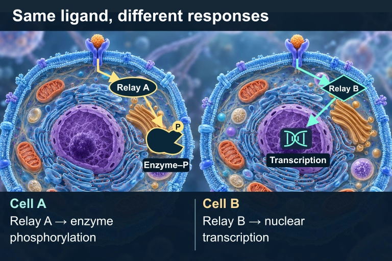
The final step produces a specific cellular response. The same signaling molecule can trigger different responses in different cell types because each cell expresses different proteins.
Gene Expression
Transcription factors are activated, turning specific genes on or off.
Enzyme Activation
Metabolic enzymes are activated or inhibited, altering cell metabolism.
Cytoskeletal Changes
The cell may change shape, move, or divide in response to the signal.
Two cells have matching ligands and receptors. In Cell A, an oval cytoplasmic Relay A points to a separate cytoplasmic Enzyme–P with a phosphate badge. In Cell B, a diamond cytoplasmic Relay B points to a DNA/transcription icon inside the nucleus.
The same compatible ligand and receptor can produce different responses because the cells express different intracellular relays and effectors.

GPCR vs. RTK

G Protein-Coupled Receptor Receptor Tyrosine Kinase
Structure 7 transmembrane helices Single transmembrane helix per monomer
Activation Activates G protein (\( \text{GTP} \)/\( \text{GDP} \) exchange) Dimerizes and auto-phosphorylates tyrosines
Second Messenger \( \text{cAMP} \), \( \text{IP}_3 \), \( \text{Ca}^{2+} \) None (\( \text{Ras} \to \text{MAP kinase cascade} \))
Speed Fast (seconds) Slower (minutes); often affects gene expression
Example Epinephrine signaling Growth factor signaling (EGF, insulin)

The Role of Second Messengers

Second messengers are small, non-protein molecules that relay signals received at the cell surface to target molecules inside the cell. They spread the signal quickly because they are small and diffuse rapidly.

\( \text{cAMP} \)
Cyclic AMP: produced by adenylyl cyclase from \( \text{ATP} \). Activates protein kinase A (PKA).
\( \text{Ca}^{2+} \)
Calcium ions: released from ER stores. Binds calmodulin to activate diverse targets.
\( \text{IP}_3 \) / \( \text{DAG} \)
Inositol trisphosphate opens \( \text{Ca}^{2+} \) channels. \( \text{DAG} \) activates protein kinase C.

Phosphorylation Cascade

Kinase 1 + P Kinase 1* (active) + P Kinase 2* (active) + P Kinase 3* (active) Response Phosphatases remove P to deactivate

How to Analyze a Pathway

1
Identify the ligand. What molecule initiates the signal? Is it a hormone, neurotransmitter, or growth factor?
2
Identify the receptor type. Is it a GPCR, RTK, ion channel, or intracellular receptor? This determines the transduction mechanism.
3
Trace the transduction steps. List each relay molecule in order. Note where amplification occurs and which second messengers are involved.
4
Determine the response. What does the cell ultimately do: change gene expression, alter metabolism, or change shape?
5
Consider termination. How is the signal turned off? Phosphatases, GTPase activity, or ligand degradation?

Example: Epinephrine Signaling

Epinephrine GPCR G protein (GTP active) Adenylyl Cyclase (activated)
\( \text{ATP} \to \text{cAMP} \)
cAMP PKA Glycogen Breakdown → Glucose release

Predict why persistent intracellular activity can outlast ligand binding.

Inactive Gαs–GDP ceases to stimulate cyclase, while hydrolysis-defective Gαs–GTP can continue stimulation after the hormone pulse. PDE-mediated cAMP degradation continues in both. The selected signal-stimulated component excludes basal cyclase activity; repeated raster panels show contextual hardware, not measured states.
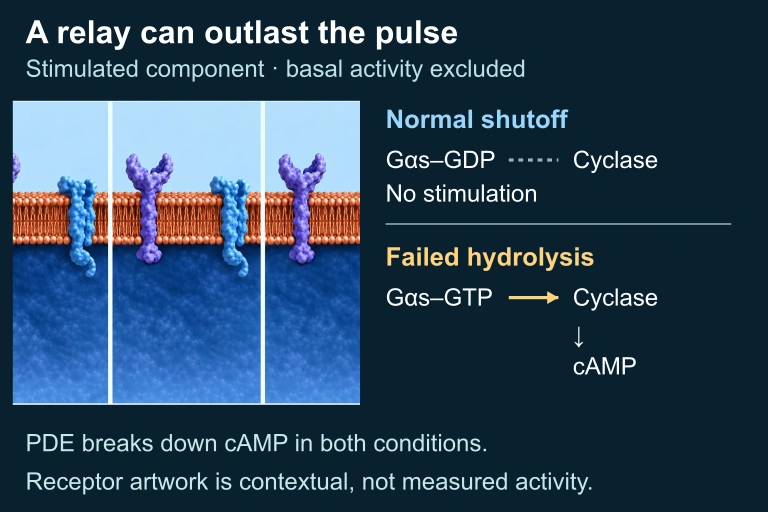
A contextual membrane illustration accompanies normal G-alpha-s GDP without stimulated cyclase activity and persistent GTP-bound G-alpha-s stimulating cyclase and cAMP formation. PDE continues to degrade cAMP in both.
Inactive Gαs–GDP ceases to stimulate cyclase, while hydrolysis-defective Gαs–GTP can continue stimulation after the hormone pulse. PDE-mediated cAMP degradation continues in both. The selected signal-stimulated component excludes basal cyclase activity; repeated raster panels show contextual hardware, not measured states.

Example: Insulin via RTK

1
Reception: Insulin binds to the receptor tyrosine kinase (RTK) on the cell surface, causing dimerization of receptor monomers.
2
Auto-phosphorylation: Each monomer phosphorylates the other's tyrosine residues, creating docking sites for relay proteins.
3
Signal relay: Ras protein is activated \( \to \) triggers the MAP kinase cascade \( \to \) phosphorylation of multiple transcription factors.
4
Response: GLUT4 vesicles fuse with membrane, allowing glucose uptake. Gene expression changes promote cell growth.

Example: Apoptosis Signaling

Apoptosis (programmed cell death) is a tightly controlled signaling process. When a cell receives a "death signal," it activates an internal self-destruct program.

Death Ligand
External signals (e.g., Fas ligand) bind to death receptors on the target cell surface.
Caspase Cascade
Initiator caspases activate executioner caspases in a proteolytic cascade.
Cell Dismantled
DNA is fragmented, organelles are destroyed, and cell shrinks into apoptotic bodies.

Common Mistakes to Avoid

✕ Ligand enters the cell
Most ligands bind extracellular receptors and do NOT cross the membrane. Only steroid hormones and other hydrophobic molecules enter directly.
✕ Confusing kinase and phosphatase
Kinases add phosphate groups (activate). Phosphatases remove them (deactivate). Mixing these up reverses the logic of the pathway.
✕ One ligand = one response
The same ligand can trigger different responses in different cell types. The response depends on which proteins the target cell expresses.

Summary of Key Pathways

GPCR Pathway RTK Pathway Apoptosis
Receptor 7-pass transmembrane Tyrosine kinase Death receptor (Fas)
Key Relay G protein, \( \text{cAMP} \) Ras, MAP kinase Caspase cascade
Second Messenger \( \text{cAMP} \), \( \text{Ca}^{2+} \) None (direct phosphorylation) None (protein cascade)
Typical Response Metabolism change Growth, differentiation Programmed cell death
Speed Seconds Minutes to hours Hours

Question 1 of 5

In signal transduction, what is the primary role of a second messenger such as \( \text{cAMP} \)?

Question 2 of 5

A mutation causes a Ras protein to remain permanently active (always bound to \( \text{GTP} \)). What is the most likely consequence?

Question 3 of 5

Epinephrine causes glycogen breakdown in liver cells but increases heart rate in cardiac cells. What best explains this difference?

Question 4 of 5

Which of the following correctly describes the function of phosphatases in a signal transduction pathway?

Question 5 of 5

A researcher applies a drug that blocks adenylyl cyclase. Which of the following effects would you predict?