Proteins
Structure and Function
Explore how amino acids build into complex macromolecules that drive nearly every process in living cells: from catalysis to structural support.
What Are Proteins?
Proteins are polymers made of amino acid monomers linked by peptide bonds. They are the most structurally and functionally diverse macromolecules in living systems, making up over 50% of the dry mass of most cells.
Anatomy of an Amino Acid
Peptide Bonds and Dehydration Synthesis
The Four Levels of Protein Structure
Visualizing Protein Folding
Sequence folds into local, global and multi-chain structure
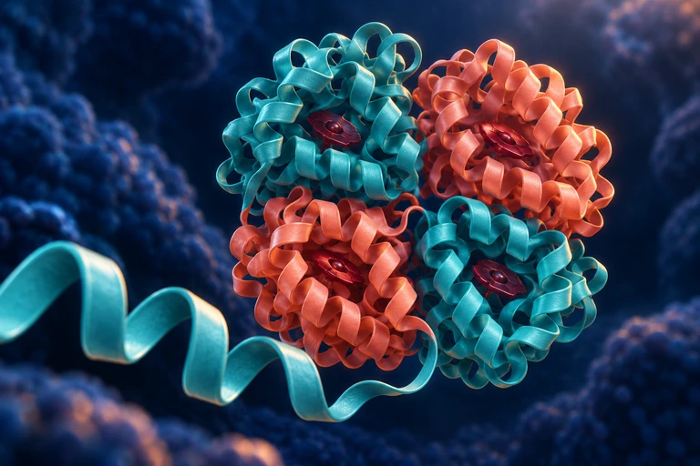
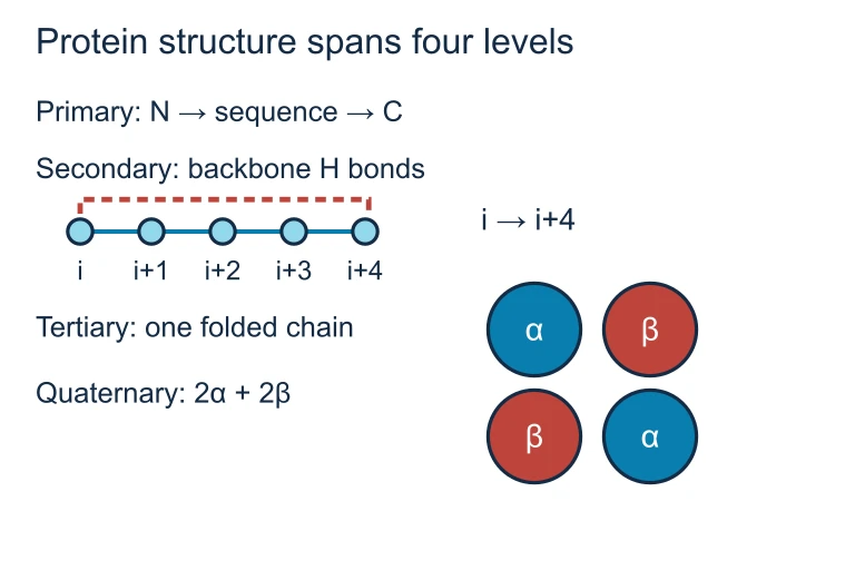
Protein organization spans amino-acid sequence, local secondary structure, a folded chain and multi-chain assemblies. An α-helix uses backbone hydrogen bonds from residue i to i+4; hemoglobin contains two α and two β chains.Protein structure spans four levels
Protein organization spans amino-acid sequence, local secondary structure, a folded chain and multi-chain assemblies. An α-helix uses backbone hydrogen bonds from residue i to i+4; hemoglobin contains two α and two β chains.Fibrous vs Globular Proteins
| Fibrous | Globular | |
|---|---|---|
| Shape | Long, rope-like strands | Compact, spherical |
| Solubility | Insoluble in water | Usually water-soluble |
| Function | Structural support | Catalysis, transport, signaling |
| Examples | Collagen, keratin | Hemoglobin, enzymes |
| Structure level | Mostly 2° (repetitive) | Complex 3° and 4° |
Major Functions of Proteins


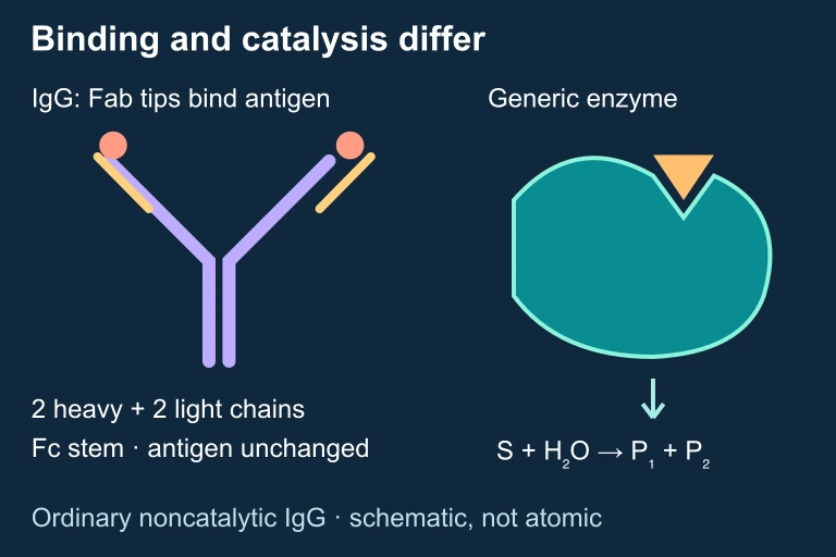
Enzymes: Lock and Key Model
Denaturation: Losing Shape
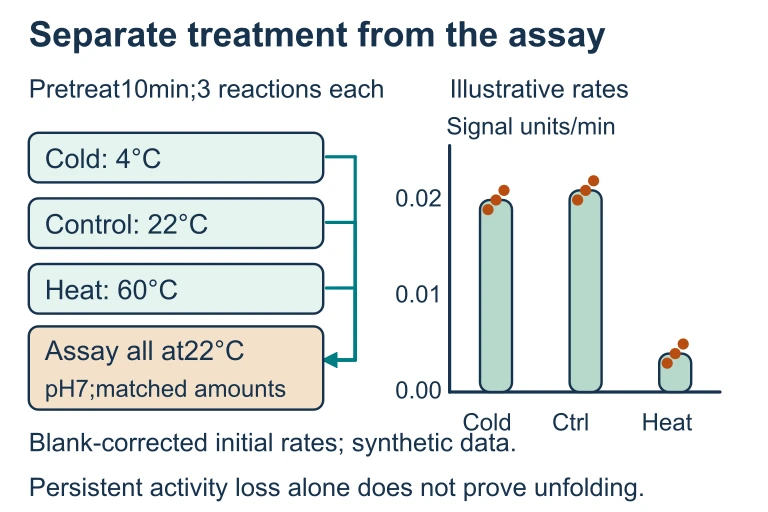
Cold, heated and recovered: an enzyme experiment with a visible control story
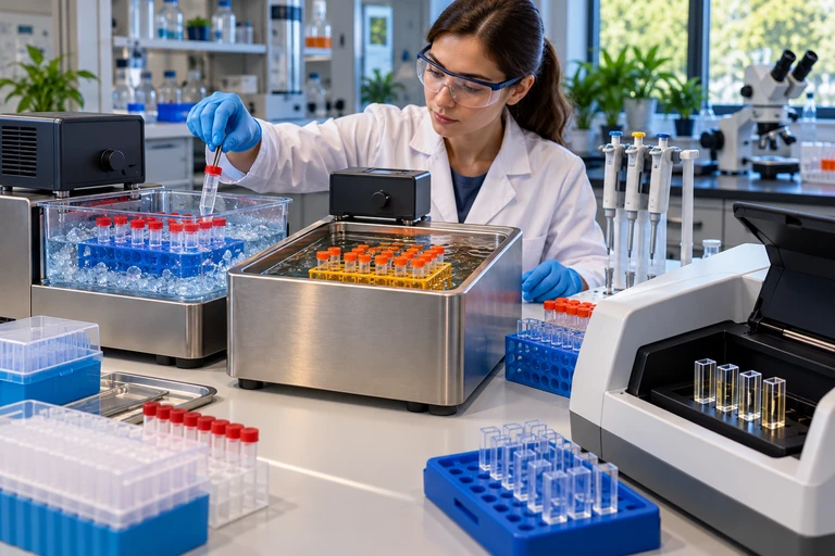
To distinguish prior temperature exposure from immediate reaction conditions, return all enzyme treatments to the same assay temperature and pH. Recovery after cooling differs from persistent activity loss after heating.Separate treatment from the assay
To distinguish prior temperature exposure from immediate reaction conditions, return all enzyme treatments to the same assay temperature and pH. Recovery after cooling differs from persistent activity loss after heating.How to Identify Structure Levels
Example: Sickle Cell Disease
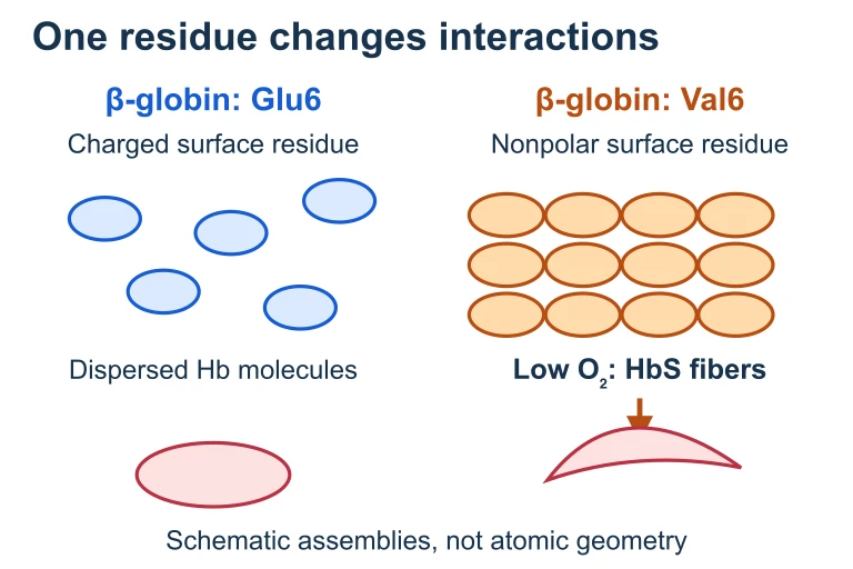
Mutation can expose a hydrophobic interaction surface
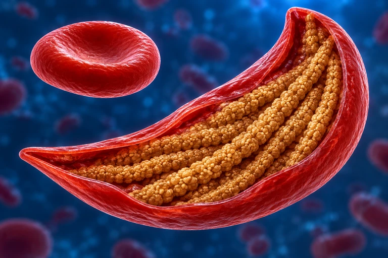
The mature β-globin Glu6→Val6 substitution replaces a charged surface residue with a nonpolar one. Under low oxygen, HbS can assemble into fibers that deform red cells.One residue changes interactions
The mature β-globin Glu6→Val6 substitution replaces a charged surface residue with a nonpolar one. Under low oxygen, HbS can assemble into fibers that deform red cells.Example: Enzyme Specificity
Question: Why can lactase break down lactose but not sucrose, even though both are disaccharides?
Example: Collagen Triple Helix
Common Mistakes on the AP Exam
Summary: Protein Structure at a Glance
| Level | What It Describes | Key Bonds | Example |
|---|---|---|---|
| Primary | Amino acid sequence | Peptide (covalent) | Insulin chain |
| Secondary | Local folds (helix, sheet) | Hydrogen (backbone) | Alpha helix in keratin |
| Tertiary | 3D shape of polypeptide | R-group interactions | Myoglobin |
| Quaternary | Multi-subunit assembly | Same as tertiary | Hemoglobin (4 subunits) |
Question 1 of 5
Which level of protein structure is directly determined by the gene sequence encoding the protein?
Question 2 of 5
A researcher heats a protein to 95°C and observes it loses its function. Which statement best describes what happened?
Question 3 of 5
Hemoglobin consists of four polypeptide subunits working together. This arrangement represents which level of protein structure?
Question 4 of 5
In sickle cell disease, the substitution of valine for glutamic acid at position 6 of the beta-globin chain causes the protein to aggregate. This demonstrates that:
Question 5 of 5
Which bond type is responsible for the formation of alpha helices and beta pleated sheets in secondary structure?