01 / 22

The Plasma
Membrane

Explore the fluid mosaic model, selective permeability, and the transport mechanisms that keep every cell alive.

AP Biology

What Is the Plasma Membrane?

The plasma membrane (cell membrane) is a thin, flexible boundary that separates a cell's interior from its external environment. It is only about 7 to 8 nm thick, yet it performs critical functions: controlling what enters and exits the cell, facilitating cell signaling, and maintaining structural integrity.

The Phospholipid Bilayer

EXTRACELLULAR FLUID CYTOPLASM Hydrophilic head Hydrophobic tails Integral protein Cholesterol

Fluid membrane architecture preserves leaflet asymmetry
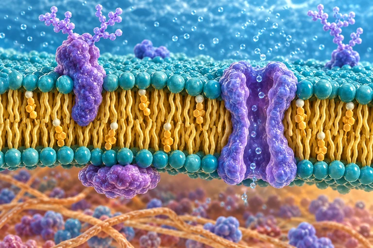
A teal-and-gold membrane cutaway has purple embedded proteins, an open channel, outward branching glycans and a protein on the lower cytoplasmic surface.Polar phospholipid heads face water on both sides of the membrane while hydrophobic tails face inward. Carbohydrate chains on glycoproteins extend from the extracellular face.

Two water-facing leaflets
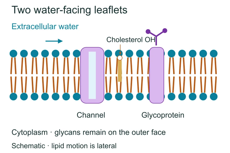
Two water-facing leaflets. Polar phospholipid heads face water on both sides of the membrane while hydrophobic tails face inward. Carbohydrate chains on glycoproteins extend from the extracellular face.Polar phospholipid heads face water on both sides of the membrane while hydrophobic tails face inward. Carbohydrate chains on glycoproteins extend from the extracellular face.

Key Components of the Membrane
Phospholipids
Form the bilayer scaffold. Their amphipathic nature creates a self-sealing barrier.
Proteins
Integral proteins span the membrane; peripheral proteins sit on the surface. Both enable transport and signaling.
Cholesterol
Wedges between phospholipid tails, maintaining fluidity across temperature changes.
Carbohydrates
Glycoproteins and glycolipids on the outer surface aid in cell recognition and immune function.
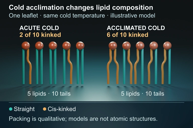
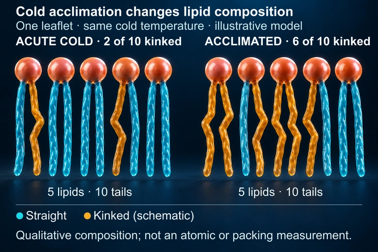
Two illustrated single leaflets each have five lipid heads and ten tails. In the acute cold example, two tails are kinked; in the acclimated cold example, six are kinked. Both are shown at the same cold temperature.
Some organisms acclimate to cold by increasing unsaturated fatty-acid content, helping maintain membrane fluidity. This qualitative composition model compares leaflets at the same cold temperature; it is not an atomic structure or packing measurement.
Authored geometry companion

The Fluid Mosaic Model

Proposed by Singer and Nicolson (1972), this model describes the membrane as a two-dimensional fluid in which proteins float like icebergs in a sea of phospholipids. The term "fluid" refers to the lateral movement of lipids and proteins, while "mosaic" describes the diverse patchwork of embedded molecules.

1
Lipids move laterally within each leaflet approximately \(10^7\) times per second.
2
Flip-flop (transverse movement between leaflets) is rare without enzyme assistance from flippases.
3
Membrane proteins drift, rotate, or are anchored by the cytoskeleton, creating functional microdomains.

Selective Permeability: The Core Rule

PHOSPHOLIPID BILAYER
\( \text{O}_2 \)
\( \text{O}_2 \)
\( \text{CO}_2 \)
Glucose CH
\( \text{Na}^+ \)
(ion) Passes freely Needs channel Blocked

Passive vs Active Transport

Passive Transport
Moves substances down their concentration gradient. No energy (ATP) required. Includes simple diffusion, facilitated diffusion, and osmosis.
Active Transport
Moves substances against their concentration gradient. Requires energy (ATP). Includes the \( \text{Na}^+/\text{K}^+ \) pump and bulk transport (endo/exocytosis).

Types of Passive Transport

Simple Diffusion
Small nonpolar molecules pass directly through the bilayer: \( \text{O}_2 \), \( \text{CO}_2 \), ethanol.
Facilitated Diffusion
Polar molecules and ions pass through channel or carrier proteins: glucose via \( \text{GLUT} \), ions via ion channels.
Osmosis
The diffusion of water across a selectively permeable membrane through aquaporins, from low solute to high solute concentration.

Osmosis and Tonicity

Cell swells Hypotonic Water enters cell Cell normal Isotonic Water balanced Cell shrinks Hypertonic Water exits cell

A plant cell presses against a wall while an animal cell changes shape
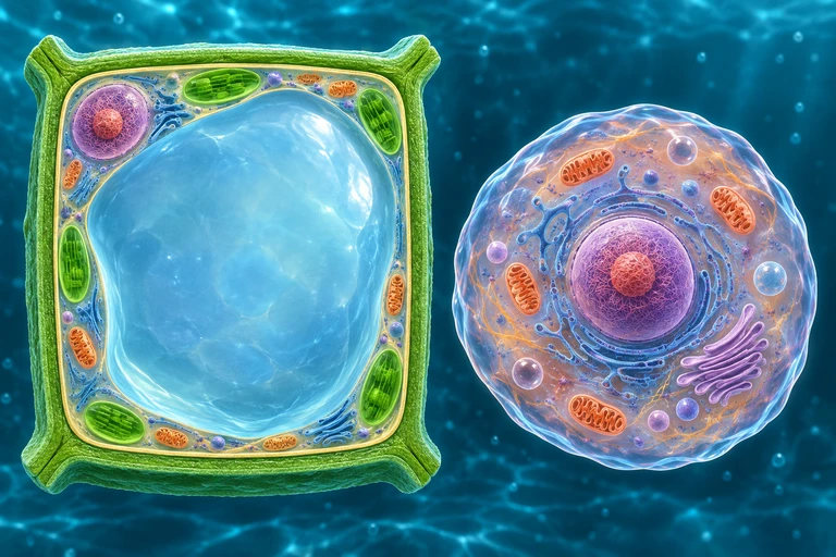
Plant wall is thick and distinct from inner membrane; large central vacuole and peripheral cytoplasm visible. Animal cell has flexible boundary and no wall. Separate exact panel: Paired plant states retain the same wall while the inner membrane is pressed outward or retracted; animal membrane has no surrounding wall.For nonpenetrating solutes, a plant wall limits expansion as water enters. Water loss retracts the protoplast from the wall during plasmolysis; animal cells lack this rigid wall.

Wall support changes the response
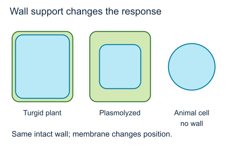
Wall support changes the response. For nonpenetrating solutes, a plant wall limits expansion as water enters. Water loss retracts the protoplast from the wall during plasmolysis; animal cells lack this rigid wall.For nonpenetrating solutes, a plant wall limits expansion as water enters. Water loss retracts the protoplast from the wall during plasmolysis; animal cells lack this rigid wall.

Active Transport: The \( \text{Na}^+/\text{K}^+ \) Pump
MEMBRANE
\( \text{Na}^+/\text{K}^+ \)
ATPase
\( \text{Na}^+ \)
\( \text{Na}^+ \)
\( \text{Na}^+ \)
\( \text{K}^+ \)
\( \text{K}^+ \)
\( \text{ATP} \rightarrow \text{ADP} + \text{P}_i \)
EXTRACELLULAR INTRACELLULAR
\( 3~\text{Na}^+ \) out
\( 2~\text{K}^+ \) in
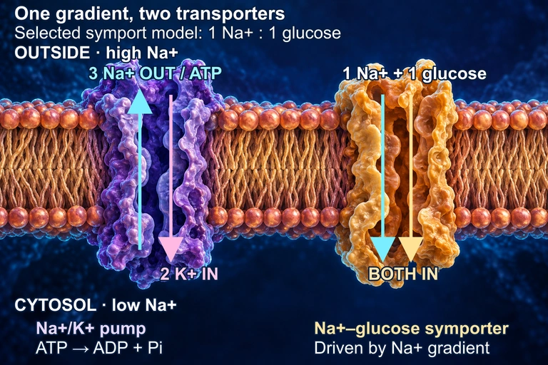
A conceptual membrane has higher sodium outside and lower sodium in the cytosol. A violet pump moves three sodium out and two potassium in per ATP. A separate gold symporter brings one sodium and one glucose inward, using the gradient. Protein forms and their juxtaposition are schematic.
In this conceptual membrane, the Na+/K+ ATPase uses one ATP to move three Na+ out and two K+ in, maintaining a sodium electrochemical gradient. A separate symporter uses that gradient to bring Na+ and glucose inward; the 1:1 ratio is a selected teaching model and varies among transporters. The cutaways do not represent constitutively open pores: both transport cycles and the protein shapes are schematic. The juxtaposition does not assert a shared membrane face in a particular tissue.

Bulk Transport: Endocytosis and Exocytosis

Endocytosis
The membrane folds inward to engulf large particles. Includes phagocytosis (cell eating), pinocytosis (cell drinking), and receptor-mediated endocytosis.
Exocytosis
Vesicles fuse with the membrane to release contents outside the cell. Used for secreting hormones, neurotransmitters, and waste removal.

How to Identify the Transport Type

1
Determine the direction. Is the substance moving from high to low concentration (down the gradient) or from low to high (against it)?
2
Check energy use. If energy (ATP) is required, it is active transport. If not, it is passive.
3
Identify the molecule. Small and nonpolar: simple diffusion. Polar or charged: facilitated diffusion or active transport. Very large: bulk transport.
4
Look for proteins. If a transport protein is involved and no ATP is used, it is facilitated diffusion. If ATP is used, it is primary active transport.

Example 1: Red Blood Cells in Solutions

A red blood cell (RBC) is placed in a 0.1% NaCl solution. Normal blood plasma is about 0.9% NaCl. What happens to the cell?

1
Compare concentrations: 0.1% solution has less solute than the cell interior (0.9%). The solution is hypotonic relative to the cell.
2
Predict water movement: Water moves by osmosis from the hypotonic solution (more water) into the cell (less water).
3
Result: The RBC swells and may lyse (burst) because animal cells lack a cell wall to resist osmotic pressure.

Example 2: Glucose Uptake in Intestinal Cells

After a meal, glucose concentration in the intestinal lumen is higher than inside epithelial cells. But the cells also absorb glucose when concentrations are equal. How?

1
High lumen glucose: Glucose enters via facilitated diffusion through \( \text{GLUT2} \) transporters (passive, down the gradient).
2
Equal concentrations: The cell uses secondary active transport (\( \text{SGLT1} \) cotransporter), coupling glucose uptake with \( \text{Na}^+ \) moving down its gradient established by the \( \text{Na}^+/\text{K}^+ \) pump.
3
Overall: The \( \text{Na}^+/\text{K}^+ \) pump uses ATP to maintain the \( \text{Na}^+ \) gradient, which indirectly drives glucose absorption: secondary active transport.

Example 3: Signal Transduction at the Membrane

A water-soluble hormone like insulin cannot cross the hydrophobic bilayer. How does the cell respond?

1
Receptor binding: Insulin binds to a receptor protein (tyrosine kinase) on the extracellular surface of the membrane.
2
Signal relay: The receptor undergoes a conformational change, activating an intracellular signaling cascade (phosphorylation events).
3
Cellular response: \( \text{GLUT4} \) vesicles fuse with the membrane via exocytosis, increasing glucose uptake. The signal never had to enter the cell.

Common Mistakes

✕ Confusing tonicity direction
Students say "water moves to the hypotonic side." Water moves toward the hypertonic side (higher solute, lower free water).
✕ Calling osmosis "active"
Osmosis is a form of passive transport. It does not require ATP, even though aquaporin channels may be involved.
✕ Saying membranes are "static"
The fluid mosaic model shows that lipids and proteins are constantly moving laterally within the bilayer, not fixed in place.

Transport Comparison

Feature Passive Active Bulk
ATP needed? No Yes Yes
Direction High → Low Low → High Into/out of cell
Protein needed? Sometimes Always Vesicles
Examples \( \text{O}_2 \), osmosis, GLUT \( \text{Na}^+/\text{K}^+ \) pump Endo/exocytosis
Molecule size Small to medium Small ions Large / macro

Question 1 of 5

A plant cell is placed in a hypertonic solution. Which outcome is most likely?

Question 2 of 5

Which molecule can pass most easily through the phospholipid bilayer without assistance?

Question 3 of 5

The \( \text{Na}^+/\text{K}^+ \) ATPase pumps \( 3~\text{Na}^+ \) out and \( 2~\text{K}^+ \) in per cycle. What is the net effect on the cell's interior charge?

Question 4 of 5

Cholesterol in the plasma membrane primarily functions to:

Question 5 of 5

A white blood cell engulfs a bacterium by wrapping its membrane around it and forming a vesicle. This process is called: