Explore how cells move substances across membranes: from passive diffusion to active transport and bulk transport.
AP Biology
What Is Membrane Transport?
Every living cell is bounded by a selectively permeable plasma membrane. This phospholipid bilayer controls which molecules enter and exit, maintaining the internal environment the cell needs to survive.
The Concentration Gradient
Two Categories: Passive vs. Active
Passive Transport
No energy (ATP) required. Molecules move down their concentration gradient: high to low.
Active Transport
Requires energy (ATP). Molecules move against their concentration gradient: low to high.
Types of Passive Transport
Simple Diffusion
Small, nonpolar molecules (\( \text{O}_2 \), \( \text{CO}_2 \)) pass directly through the lipid bilayer.
Facilitated Diffusion
Polar molecules and ions use channel or carrier proteins to cross. Still no ATP needed.
Osmosis
The diffusion of water across a selectively permeable membrane via aquaporins.
Diffusion Across the Membrane
Osmosis and Tonicity
Hypotonic
Solute lower outside. Water enters cell. Cell swells (lysis in animal cells).
Isotonic
Solute equal. No net water movement. Cell stays normal.
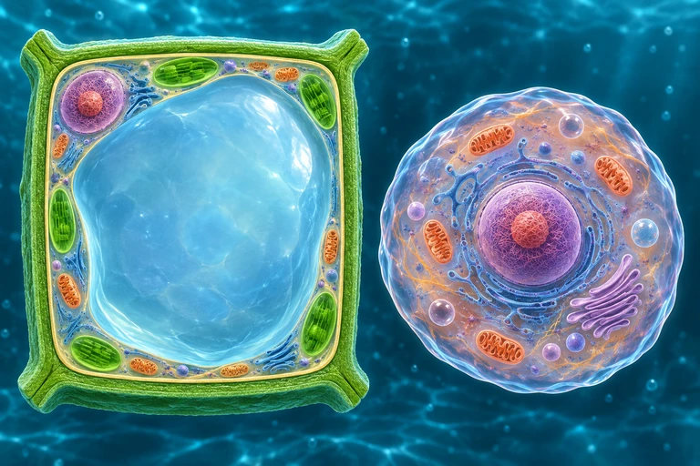
A plant cell presses against a wall while an animal cell changes shape
For nonpenetrating solutes, a plant wall limits expansion as water enters. Water loss retracts the protoplast from the wall during plasmolysis; animal cells lack this rigid wall.
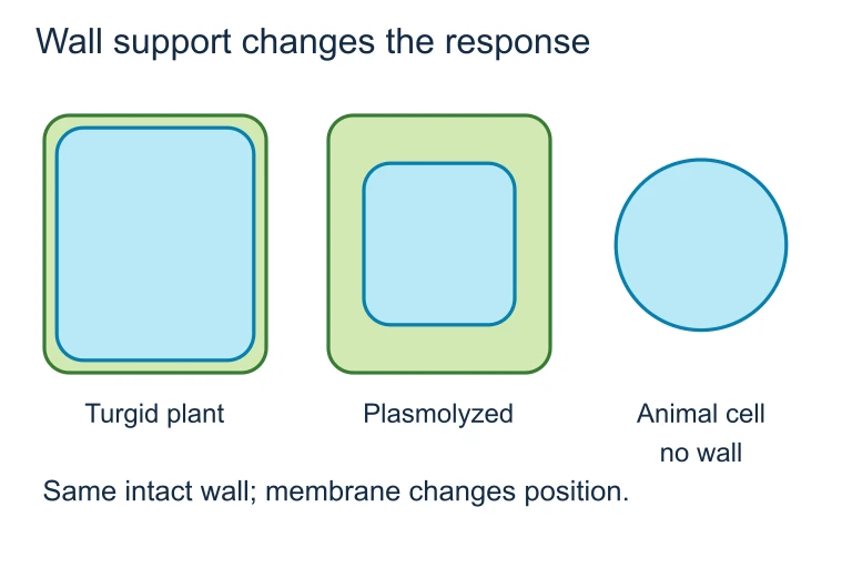
Wall support changes the response
For nonpenetrating solutes, a plant wall limits expansion as water enters. Water loss retracts the protoplast from the wall during plasmolysis; animal cells lack this rigid wall.
Active Transport: Against the Gradient
The \( \text{Na}^+/\text{K}^+ \) ATPase Pump
1
\( 3 \ \text{Na}^+ \) ions bind to the pump on the cytoplasmic side.
2
ATP is hydrolyzed to \( \text{ADP} + \text{P}_i \), phosphorylating the pump and changing its shape.
3
\( 3 \ \text{Na}^+ \) are released to the extracellular side.
4
\( 2 \ \text{K}^+ \) ions bind on the extracellular side.
5
Phosphate is released, pump returns to original shape, \( 2 \ \text{K}^+ \) enter the cell.
Bulk Transport: Endocytosis & Exocytosis
Endocytosis
Membrane folds inward to bring large molecules into the cell inside a vesicle.
Exocytosis
Vesicle fuses with membrane to release contents outside the cell.
Phagocytosis
"Cell eating": engulfs large particles (e.g., white blood cells engulfing bacteria).
Pinocytosis
"Cell drinking": takes in dissolved molecules in small vesicles.
Receptor-Mediated
Specific ligands bind receptors, triggering coated-pit endocytosis (e.g., cholesterol via LDL).
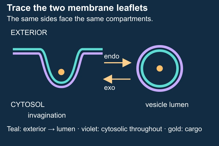
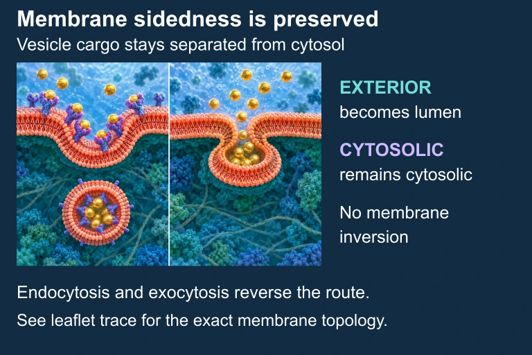
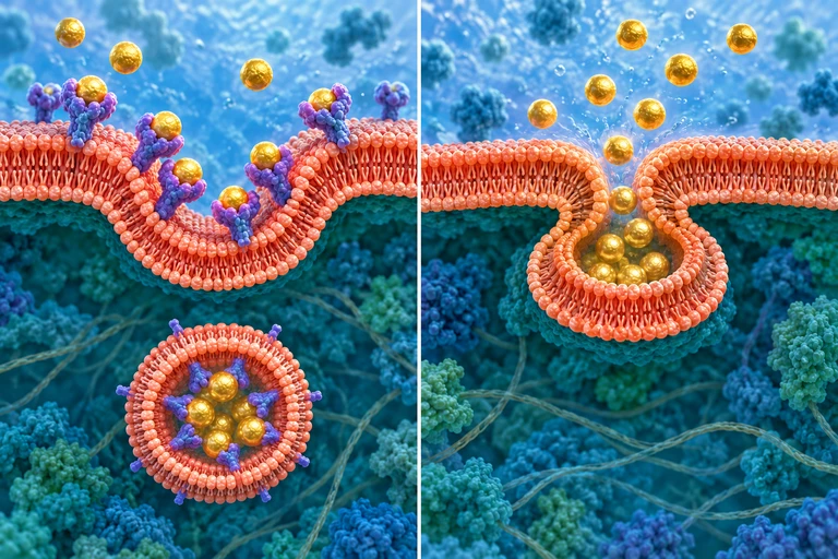
Trace the two membrane leaflets through vesicle formation and fusion.Vesicle budding and fusion preserve which membrane surface faces cytosol and which faces the lumen or exterior. Illustration is schematic where scale or molecular structure is simplified.
Vesicular transport preserves topology
Endocytosis encloses extracellular cargo in a vesicle lumen; exocytosis opens that lumen to the extracellular space. Membrane fusion and budding preserve which membrane side faces the cargo.
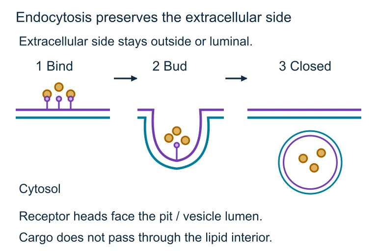
Endocytosis preserves the extracellular side
Endocytosis encloses extracellular cargo in a vesicle lumen; exocytosis opens that lumen to the extracellular space. Membrane fusion and budding preserve which membrane side faces the cargo.
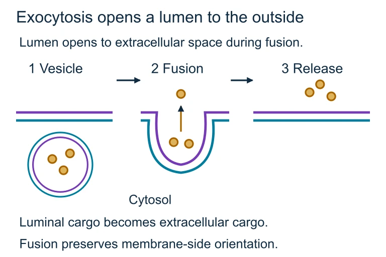
Exocytosis opens a lumen to the outside
During exocytosis, the vesicle lumen opens to the extracellular space while membrane-side orientation is preserved.
Check the direction. Is the molecule moving from high to low concentration (down gradient) or low to high (against gradient)?
2
Check for energy. Is ATP being consumed? If yes, it is active transport.
3
Check the molecule's properties. Small and nonpolar? Likely simple diffusion. Large, polar, or charged? Needs a protein.
4
Check for vesicles. Is the membrane wrapping around something? That is bulk transport (endocytosis/exocytosis).
Example 1: \( \text{O}_2 \) Entering a Cell
Scenario: A red blood cell passes through the lung capillaries. The partial pressure of \( \text{O}_2 \) is higher in the alveoli than inside the red blood cell.
Example 2: Glucose Uptake
Scenario: After a meal, blood glucose is high. Glucose must enter muscle cells where its concentration is lower.
Example 3: Nerve Cells and the \( \text{Na}^+/\text{K}^+ \) Pump
Scenario: A neuron must maintain a resting membrane potential of approximately \( -70 \ \text{mV} \). \( \text{Na}^+ \) concentration is already higher outside the cell; \( \text{K}^+ \) is higher inside.
\( 3 \ \text{Na}^+ \) pumped out
Against the gradient (already high outside). Costs 1 ATP per cycle.
\( 2 \ \text{K}^+ \) pumped in
Against the gradient (already high inside). Same ATP hydrolysis.
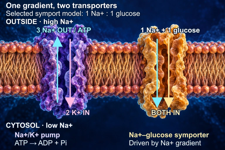
In this conceptual membrane, the Na+/K+ ATPase uses one ATP to move three Na+ out and two K+ in, maintaining a sodium electrochemical gradient. A separate symporter uses that gradient to bring Na+ and glucose inward; the 1:1 ratio is a selected teaching model and varies among transporters. The cutaways do not represent constitutively open pores: both transport cycles and the protein shapes are schematic. The juxtaposition does not assert a shared membrane face in a particular tissue.
Common Mistakes
✕ Confusing direction of osmosis
Water moves toward higher solute concentration, not away from it. Think: water follows solute.
✕ Calling facilitated diffusion "active"
Using a protein does not mean energy is needed. Facilitated diffusion still goes down the gradient: no ATP.
✕ Forgetting bulk transport
Endocytosis and exocytosis are also active (ATP-dependent) processes. Students often only list pumps as "active."
Summary: All Transport Types
Type
Energy?
Protein?
Example
Simple Diffusion
No
No
\( \text{O}_2 \), \( \text{CO}_2 \)
Facilitated Diffusion
No
Yes
Glucose, ions
Osmosis
No
Aquaporin
\( \text{H}_2\text{O} \)
Primary Active
Yes (ATP)
Pump
\( \text{Na}^+/\text{K}^+ \) ATPase
Secondary Active
Indirect
Co-transporter
SGLT (glucose/\( \text{Na}^+ \))
Endocytosis
Yes
Vesicle
Phagocytosis
Exocytosis
Yes
Vesicle
Neurotransmitter release
Question 1 of 5
A plant cell is placed in a hypertonic solution. Which of the following will occur?
In a hypertonic solution, solute concentration is higher outside. Water moves out of the cell, causing the plasma membrane to pull away from the cell wall: plasmolysis.
Question 2 of 5
Glucose enters a cell via a GLUT transporter, moving from high to low concentration. This is an example of:
Glucose moves down its concentration gradient (no ATP), but it requires a carrier protein (GLUT). This is facilitated diffusion: passive transport using a membrane protein.
Question 3 of 5
The \( \text{Na}^+/\text{K}^+ \) ATPase pump moves \( 3 \ \text{Na}^+ \) out and \( 2 \ \text{K}^+ \) in per ATP hydrolyzed. Why is this pump considered electrogenic?
The pump exports 3 positive charges (\( \text{Na}^+ \)) but imports only 2 (\( \text{K}^+ \)), producing a net loss of positive charge inside. This unequal charge transfer makes the pump electrogenic, contributing to the negative resting membrane potential.
Question 4 of 5
A white blood cell engulfs a bacterium by wrapping its membrane around it and forming a vesicle. This process is called:
Phagocytosis ("cell eating") is the engulfment of large particles or organisms. The membrane extends pseudopods around the bacterium, forming a phagosome that fuses with a lysosome for digestion.
Question 5 of 5
A researcher adds a metabolic poison that stops all ATP production in a cell. Which transport process would continue functioning?
Osmosis is passive transport: it requires no ATP. Water moves down its concentration gradient through aquaporins regardless of the cell's energy supply. All other options (pump, phagocytosis, exocytosis) require ATP and would stop.
 For nonpenetrating solutes, a plant wall limits expansion as water enters. Water loss retracts the protoplast from the wall during plasmolysis; animal cells lack this rigid wall.
 For nonpenetrating solutes, a plant wall limits expansion as water enters. Water loss retracts the protoplast from the wall during plasmolysis; animal cells lack this rigid wall.
 Endocytosis encloses extracellular cargo in a vesicle lumen; exocytosis opens that lumen to the extracellular space. Membrane fusion and budding preserve which membrane side faces the cargo.
 Endocytosis encloses extracellular cargo in a vesicle lumen; exocytosis opens that lumen to the extracellular space. Membrane fusion and budding preserve which membrane side faces the cargo.
 During exocytosis, the vesicle lumen opens to the extracellular space while membrane-side orientation is preserved.