01 / 22

Introduction to
Signal Transduction

Explore how cells receive, process, and respond to chemical signals: the three-stage pathway that governs everything from growth to immune defense.

AP Biology

What Is Cell Communication?

Cells do not exist in isolation. They constantly send and receive chemical messages that coordinate activities throughout an organism: controlling growth, triggering immune responses, and maintaining homeostasis.

Think of it like a relay race: a message is received at the cell surface, passed along through a series of molecular handoffs, and finally triggers an action inside the cell.

Key Vocabulary

Ligand
A signaling molecule (hormone, neurotransmitter, or growth factor) that binds to a specific receptor.
Receptor
A protein on or in the target cell that specifically recognizes and binds the ligand, initiating a response.
Second Messenger
A small intracellular molecule (e.g., \( \text{cAMP} \), \( \text{Ca}^{2+} \)) that relays and amplifies the signal inside the cell.

Why Does It Matter?

Signal transduction is essential for multicellular life. Without it, cells could not coordinate development, respond to danger, or maintain internal balance.

Growth
Growth factors signal cells to divide at the right time and place during development.
Immunity
Immune cells detect foreign antigens and trigger cascades that destroy pathogens.
Homeostasis
Hormones like insulin signal cells to absorb glucose, keeping blood sugar stable.

The Three Stages of Signal Transduction

1. Reception Ligand binds receptor 2. Transduction Relay molecules cascade 3. Response Cell activity changes Signal Transduction Pathway

Stage 1: Reception

PLASMA MEMBRANE Receptor Ligand Intracellular Domain Conformational change Extracellular Intracellular

Types of Receptors

G-Protein Coupled
7-transmembrane domain proteins that activate G-proteins on the cytoplasmic side. Most common receptor type in humans.
Receptor Tyrosine Kinase
Dimerize upon ligand binding and cross-phosphorylate tyrosine residues, triggering multiple downstream pathways simultaneously.
Ion Channel
Ligand-gated channels that open upon binding, allowing ions (like \( \text{Na}^+ \) or \( \text{Ca}^{2+} \)) to flow in or out rapidly.
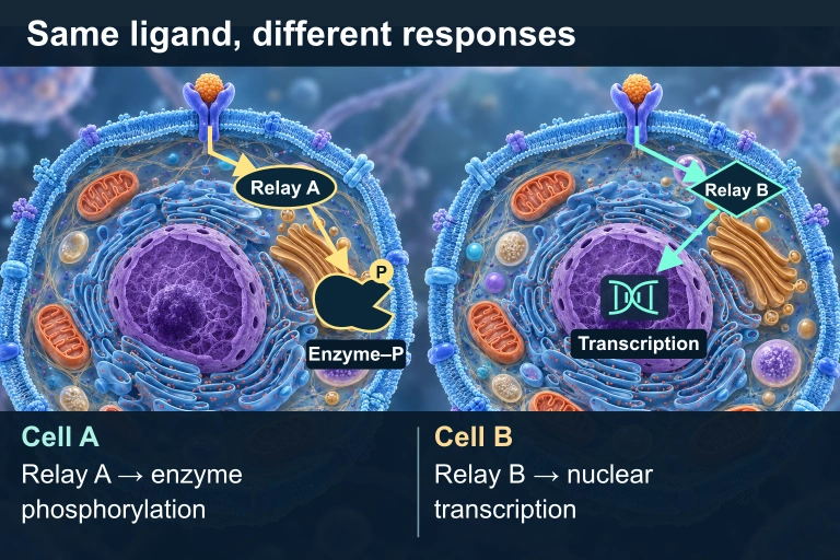
Two cells have matching ligands and receptors. In Cell A, an oval cytoplasmic Relay A points to a separate cytoplasmic Enzyme–P with a phosphate badge. In Cell B, a diamond cytoplasmic Relay B points to a DNA/transcription icon inside the nucleus.
The same compatible ligand and receptor can produce different responses because the cells express different intracellular relays and effectors.

Distance and route of cell communication
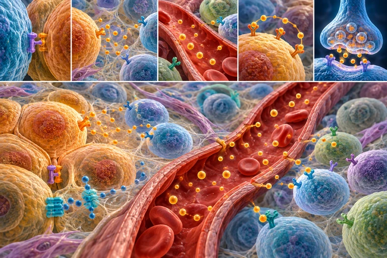
Five tissue-route details plus a larger tissue view show contact, local signaling, blood transport, self-response and synaptic release. Ligand identity and receptor selectivity require the exact layer.Cell-surface receptors connect extracellular ligand binding to intracellular responses. GPCRs, receptor tyrosine kinases and ligand-gated ion channels use distinct architectures.

Three membrane-receptor architectures
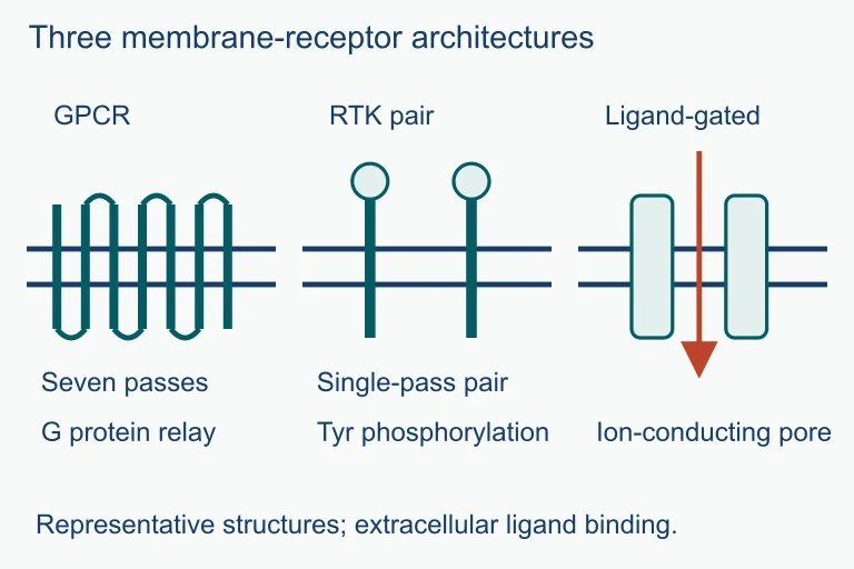
Three membrane-receptor architectures. GPCRs relay signals through G proteins, receptor tyrosine kinases use phosphorylation, and ligand-gated channels open an ion-conducting pore.GPCRs relay signals through G proteins, receptor tyrosine kinases use phosphorylation, and ligand-gated channels open an ion-conducting pore.

Stage 2: Transduction

Receptor (activated) Relay 1 (kinase) Relay 2 (kinase) Relay 3 (activated) P P P Signal Amplification x10 x100 x1000 Each step multiplies the signal exponentially

Second Messengers

Second messengers are small, non-protein molecules that spread the signal rapidly throughout the cell. They act as molecular amplifiers between the receptor and the cellular response.

\( \text{cAMP} \)
Cyclic adenosine monophosphate. Produced by adenylyl cyclase from \( \text{ATP} \). Activates protein kinase A (PKA).
\( \text{Ca}^{2+} \) Ions
Released from the ER into the cytoplasm. Triggers muscle contraction, exocytosis, and enzyme activation.
\( \text{IP}_3 \) / \( \text{DAG} \)
Inositol trisphosphate and diacylglycerol. \( \text{IP}_3 \) releases \( \text{Ca}^{2+} \) from ER; \( \text{DAG} \) activates protein kinase C.

Stage 3: Response

The final stage converts the transduced signal into a specific cellular action. Different cell types can respond differently to the same signal.

1
Gene expression changes: transcription factors activate or repress specific genes (e.g., cell growth genes).
2
Enzyme activation: metabolic enzymes are switched on or off, altering cellular metabolism.
3
Cytoskeletal rearrangement: the cell changes shape, moves, or divides.
4
Apoptosis: programmed cell death is triggered when damaged or infected cells receive "death signals."

Paracrine vs Endocrine Signaling

Paracrine Endocrine
Distance Short range (nearby cells) Long range (via bloodstream)
Signal type Local regulators (growth factors) Hormones
Speed Fast (seconds to minutes) Slower (minutes to hours)
Example Neurotransmitters at synapses Insulin from pancreas
Specificity Targets cells in local area Targets cells with matching receptors anywhere

How to Analyze a Signaling Pathway

1
Identify the ligand: What molecule is the signal? Is it a hormone, growth factor, or neurotransmitter?
2
Identify the receptor type: Is it a GPCR, RTK, ion channel, or intracellular receptor? This determines the transduction mechanism.
3
Trace the transduction steps: What relay molecules and second messengers are involved? Where does amplification occur?
4
Determine the response: What changes in the cell? Gene expression, enzyme activity, or cell movement?
5
Consider termination: How is the signal turned off? Phosphatases remove phosphate groups; \( \text{cAMP} \) is broken down by phosphodiesterase.

Example: Epinephrine Pathway

Epinephrine (ligand) GPCR (receptor) G-protein (activates) Adenylyl cyclase (enzyme)
\( \text{cAMP} \)
(2nd msg) Glycogen breakdown
\( \text{ATP} \)

Example: Insulin and RTK

MEMBRANE Dimerization Insulin P P Ras-MAPK Cell growth PI3K-Akt Survival GLUT4 Glucose uptake RTKs activate multiple pathways simultaneously

Signal Termination

Cells must be able to turn off signals when the stimulus is no longer present. Without termination, signals would persist indefinitely, leading to diseases like cancer.
1
Phosphatases remove phosphate groups from relay proteins, deactivating them: the reverse of kinase action.
2
Phosphodiesterase degrades \( \text{cAMP} \) into \( \text{AMP} \), stopping the second messenger signal.
3
Receptor internalization: the receptor-ligand complex is taken into the cell via endocytosis and degraded.
4
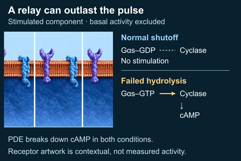
GTPase activity: G-proteins hydrolyze \( \text{GTP} \) to \( \text{GDP} \), returning themselves to an inactive state.
A contextual membrane illustration accompanies normal G-alpha-s GDP without stimulated cyclase activity and persistent GTP-bound G-alpha-s stimulating cyclase and cAMP formation. PDE continues to degrade cAMP in both.
Inactive Gαs–GDP ceases to stimulate cyclase, while hydrolysis-defective Gαs–GTP can continue stimulation after the hormone pulse. PDE-mediated cAMP degradation continues in both. The selected signal-stimulated component excludes basal cyclase activity; repeated raster panels show contextual hardware, not measured states.

Common Mistakes

✕ Ligand enters cell
Most ligands do not enter the cell. They bind extracellular receptors and trigger intracellular changes without crossing the membrane. Only hydrophobic signals (steroids) cross.
✕ One signal = one response
The same ligand can produce different responses in different cell types. Epinephrine dilates blood vessels in muscles but constricts them in the gut.
✕ Confusing kinases and phosphatases
Kinases add phosphate groups (activate). Phosphatases remove them (deactivate). They are opposing forces in the signaling balance.

Summary Comparison

Feature GPCR Pathway RTK Pathway Ion Channel
Receptor type 7-TM domain Enzyme-linked dimer Ligand-gated pore
Key intermediary G-protein, \( \text{cAMP} \) Ras, MAPK cascade Ion flux (\( \text{Ca}^{2+} \), \( \text{Na}^+ \))
Amplification High (\( \text{cAMP} \) cascade) High (kinase cascade) Low (direct ion entry)
Speed Moderate Moderate Very fast
Example ligand Epinephrine Insulin, EGF Acetylcholine

Question 1 of 5

Which of the following correctly lists the three stages of cell signaling in order?

Question 2 of 5

A scientist treats cells with a drug that inhibits phosphodiesterase. What is the most likely effect?

Question 3 of 5

Steroid hormones like testosterone differ from water-soluble signals because they:

Question 4 of 5

A mutation causes a Ras protein to be unable to hydrolyze \( \text{GTP} \). What is the most likely consequence?

Question 5 of 5

Epinephrine causes glycogen breakdown in liver cells but causes smooth muscle relaxation in bronchial cells. Which concept best explains this?