01 / 22

Cell
Communication

Explore how cells send, receive, and respond to signals: from ligand-receptor binding to intracellular signal transduction cascades.

AP Biology

Why Do Cells Communicate?

In multicellular organisms, trillions of cells must coordinate their behavior. Cell communication allows organisms to grow, respond to the environment, maintain homeostasis, and fight disease. Even single-celled organisms communicate: bacteria use quorum sensing to detect population density.

The Three Stages of Cell Signaling

1. Reception
A signaling molecule (ligand) binds to a receptor protein on or inside the target cell.
2. Transduction
The signal is converted into a form that can bring about a cellular response, often via a relay cascade.
3. Response
The cell changes its behavior: gene expression, enzyme activity, cell shape, or division.

Types of Signaling

Endocrine
Hormones travel through the bloodstream to distant target cells (long range).
Paracrine
Signals act on nearby cells via local regulators (short range).
Autocrine
A cell signals itself, reinforcing its own response.
Direct Contact
Cell-cell junctions or surface molecules allow direct signaling between adjacent cells.

Distance and route of cell communication
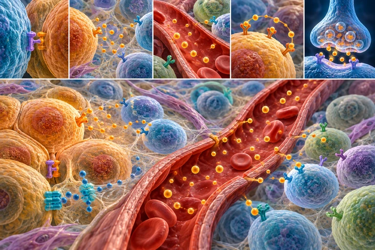
Five tissue-route details plus a larger tissue view show contact, local signaling, blood transport, self-response and synaptic release. Ligand identity and receptor selectivity require the exact layer.Signaling range describes how a ligand reaches a cell. A matching receptor determines whether that cell can respond to the signal.

Signals travel; receptors select
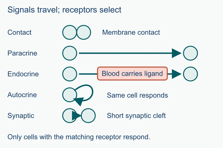
Signals travel; receptors select. Signaling range describes how a ligand reaches a cell. A matching receptor determines whether that cell can respond to the signal.Signaling range describes how a ligand reaches a cell. A matching receptor determines whether that cell can respond to the signal.

Reception: Ligand Meets Receptor

Cell Membrane Receptor Protein Intracellular L Ligand Specific binding site EXTRACELLULAR INTRACELLULAR Conformational Change Relay molecule activated

Types of Receptors

G Protein-Coupled
Seven-pass transmembrane receptor. Ligand binding activates a G protein, which activates an enzyme (e.g., adenylyl cyclase).
Receptor Tyrosine Kinase
Ligand binding causes dimerization and autophosphorylation. Activates multiple relay proteins at once.
Ion Channel Receptor
Ligand binding opens or closes a channel, allowing ions to flow across the membrane. Fast response.
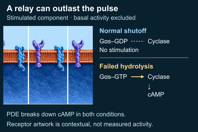
A contextual membrane illustration accompanies normal G-alpha-s GDP without stimulated cyclase activity and persistent GTP-bound G-alpha-s stimulating cyclase and cAMP formation. PDE continues to degrade cAMP in both.
Inactive Gαs–GDP ceases to stimulate cyclase, while hydrolysis-defective Gαs–GTP can continue stimulation after the hormone pulse. PDE-mediated cAMP degradation continues in both. The selected signal-stimulated component excludes basal cyclase activity; repeated raster panels show contextual hardware, not measured states.
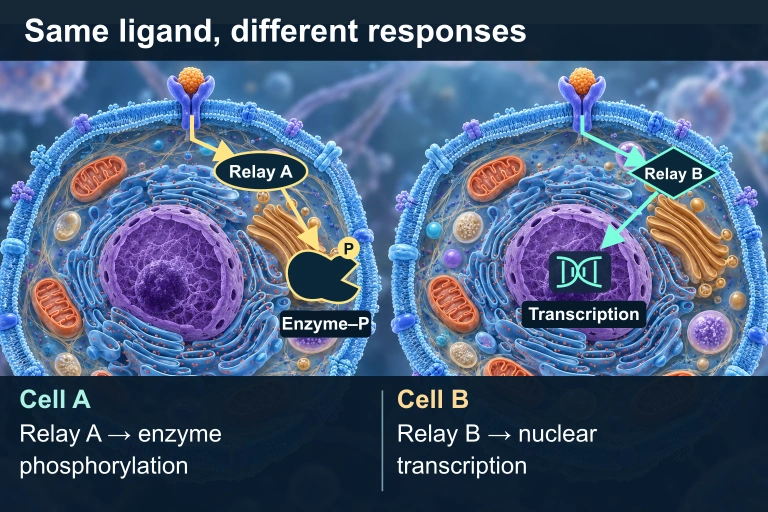
Two cells have matching ligands and receptors. In Cell A, an oval cytoplasmic Relay A points to a separate cytoplasmic Enzyme–P with a phosphate badge. In Cell B, a diamond cytoplasmic Relay B points to a DNA/transcription icon inside the nucleus.
The same compatible ligand and receptor can produce different responses because the cells express different intracellular relays and effectors.

Signal Transduction Cascade

Ligand binds receptor G protein activated Adenylyl cyclase activated cAMP produced (second messenger) Protein Kinase A activated Phosphorylation cascade Cellular Response gene expression / enzyme Signal Amplification: each step multiplies the signal 1 ligand → many activated enzymes → massive response

Second Messengers: cAMP and \( \text{Ca}^{2+} \)

Second messengers are small, non-protein molecules that relay signals from the receptor to target molecules inside the cell. They spread the signal rapidly and enable signal amplification.

cAMP
Produced from ATP by adenylyl cyclase. Activates Protein Kinase A. Degraded by phosphodiesterase.
\( \text{Ca}^{2+} \) ions
Released from the ER via \( \text{IP}_3 \)-gated channels. Involved in muscle contraction, secretion, and cell division.

Phosphorylation Cascades

1
Kinases add phosphate groups to proteins, changing their shape and activity (activation or inactivation).
2
Each activated kinase phosphorylates the next kinase in the cascade, amplifying the signal at each step.
3
Phosphatases remove phosphate groups, turning the signal off and resetting the pathway.

Paracrine vs Endocrine Signaling

Paracrine Endocrine
Range Short (nearby cells) Long (via bloodstream)
Speed Fast (seconds) Slower (minutes to hours)
Signal molecule Local regulators (growth factors) Hormones (insulin, epinephrine)
Specificity Nearby cells with receptors Distant cells with receptors
Duration Brief, localized Sustained, widespread

Cellular Responses

Gene Expression
Activated transcription factors bind DNA promoters, turning genes on or off.
Enzyme Activation
Phosphorylation activates metabolic enzymes (e.g., glycogen phosphorylase).
Cytoskeletal Change
Signals rearrange the cytoskeleton, causing cell movement or shape change.

How to Trace a Signaling Pathway

1
Identify the signal: What is the ligand? Is it hydrophobic (enters cell) or hydrophilic (binds surface receptor)?
2
Identify the receptor: GPCR, RTK, ion channel, or intracellular? This determines the transduction mechanism.
3
Follow the cascade: What second messengers are involved? cAMP, \( \text{Ca}^{2+} \), \( \text{IP}_3 \)?
4
Find the effector: What final protein or gene is activated/inhibited?
5
Predict the response: How does the cell change? Gene expression, enzyme activity, movement, or apoptosis?

Example: Epinephrine Signaling

Epinephrine GPCR G protein Adenylyl cyclase cAMP PKA Glycogen breakdown Each step amplifies the signal: 1 epinephrine → 100s of cAMP → 10,000s of glucose released

Example: Insulin and RTK Signaling

1
Insulin binds to the Receptor Tyrosine Kinase (RTK) on muscle and fat cells.
2
Two receptor subunits dimerize and cross-phosphorylate each other (autophosphorylation).
3
Phosphorylated tyrosines recruit relay proteins that activate multiple downstream pathways simultaneously.
4
One pathway triggers vesicle fusion: GLUT4 glucose transporters are inserted into the membrane.
5
Response: Glucose is rapidly absorbed from the blood, lowering blood sugar levels.

Example: Apoptosis Signaling

Death Ligand Death Receptor Adaptor Proteins Initiator Caspases Executioner Caspases Cell death (apoptosis) DNA fragmented Organelles degraded Cell shrinks

Common Mistakes

✕ Ligand enters the cell
Most ligands (hydrophilic) bind surface receptors and never enter the cell. Only steroid hormones and other hydrophobic signals cross the membrane.
✕ Signal = response
Reception, transduction, and response are distinct stages. The ligand does not directly cause the response: it triggers a cascade that does.
✕ One signal, one response
The same signal can produce different responses in different cell types. Epinephrine causes glycogen breakdown in liver cells but muscle contraction in heart cells.

Summary: Receptor Comparison

GPCR RTK Ion Channel
Structure 7-pass transmembrane Single-pass, dimerizes Multi-subunit channel
Signal mechanism G protein → enzyme Autophosphorylation Ion flow
Key pathway cAMP, \( \text{IP}_3 \) Ras → MAPK cascade \( \text{Ca}^{2+} \), \( \text{Na}^{+} \) influx
Speed Moderate Moderate Very fast
Example Epinephrine Insulin, growth factors Acetylcholine at synapse

Question 1 of 5

Which of the following correctly lists the three stages of cell signaling in order?

Question 2 of 5

A researcher applies a drug that inhibits phosphodiesterase. What is the most likely effect on the cell?

Question 3 of 5

Steroid hormones differ from peptide hormones in that steroids:

Question 4 of 5

Signal amplification in a GPCR pathway occurs because:

Question 5 of 5

Epinephrine causes glycogen breakdown in liver cells but causes increased heart rate in cardiac cells. This difference is best explained by: