
SECTION 1
Historical Context & Motivation
After Watson and Crick published the double-helix structure of DNA in 1953, a central mystery remained: how does the information encoded in a linear sequence of nucleotides produce the astonishing diversity of proteins that carry out virtually every cellular function? The concept of translation — the ribosome-mediated synthesis of a polypeptide from an mRNA template — emerged from decades of biochemical detective work that connected nucleic acid sequences to amino acid sequences. Understanding this process was essential because proteins are the ultimate effectors of genetic information, catalyzing metabolic reactions, providing structural support, and mediating signal transduction.
1958 • The Central Dogma
1961 • Cracking the Genetic Code
1964 • tRNA Adaptor Confirmed
1966 • Complete Codon Table
2000 • Ribosome Crystal Structure
These discoveries raised a deeper question still relevant for the AP exam: how do the molecular players — mRNA, tRNA, ribosomes, and translation factors — coordinate with precision to convert a nucleotide sequence into a correctly folded, functional protein? The sections that follow address this question systematically.
SECTION 2
Core Principles of Translation
Translation is the process by which ribosomes read the nucleotide sequence of messenger RNA (mRNA) in triplet units called codons and assemble amino acids into a polypeptide chain. The specificity of this process depends on transfer RNA (tRNA) molecules, each carrying a specific amino acid and bearing an anticodon complementary to the mRNA codon. Several foundational principles govern translation across all domains of life.
Codon–Anticodon Recognition
Aminoacyl-tRNA Synthetases
Ribosome as a Ribozyme
Reading Frame & Start/Stop Codons
Energy Cost
SECTION 3
Visual Overview of Translation
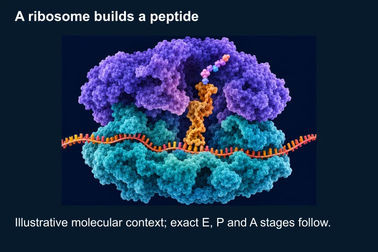
In the diagram above, note how the A (aminoacyl) site accepts incoming charged tRNAs, the P (peptidyl) site holds the tRNA linked to the growing polypeptide, and the E (exit) site releases the now-uncharged tRNA back into the cytoplasm for recycling. During each elongation cycle the ribosome translocates one codon (three nucleotides) in the 5′→3′ direction along the mRNA. Peptide bond formation between the carboxyl group of the P-site amino acid and the amino group of the A-site amino acid is catalyzed by the peptidyl transferase activity of the large subunit's rRNA.
SECTION 4
Mechanism: The Three Stages of Translation
Stage 1 — Initiation
In eukaryotes, initiation begins when eukaryotic initiation factors (eIFs) assemble a pre-initiation complex. The small ribosomal subunit (40S) associates with the initiator tRNA (Met-tRNAi) and scans the mRNA from the 5′ cap until it encounters the first AUG start codon in a favorable Kozak sequence context. The large subunit (60S) then joins, forming the complete 80S ribosome with the initiator tRNA seated in the P site. In prokaryotes, the 30S subunit recognizes the Shine-Dalgarno sequence upstream of the AUG, and the initiator amino acid is formyl-methionine (fMet). GTP hydrolysis by IF-2 (or eIF-2 in eukaryotes) powers subunit joining.
Stage 2 — Elongation
Elongation is a repetitive three-step cycle. First, codon recognition occurs when an aminoacyl-tRNA, escorted by EF-Tu·GTP (EF-1α in eukaryotes), enters the A site. If the anticodon–codon match is correct, GTP is hydrolyzed and EF-Tu releases the tRNA. Second, peptide bond formation occurs as the rRNA of the large subunit catalyzes transfer of the polypeptide from the P-site tRNA to the amino acid on the A-site tRNA. Third, translocation is driven by EF-G·GTP hydrolysis (EF-2 in eukaryotes), shifting the ribosome one codon downstream: the deacylated tRNA moves from P to E, the peptidyl-tRNA moves from A to P, and the A site is vacated for the next aminoacyl-tRNA. A single ribosome adds roughly 15–20 amino acids per second in prokaryotes and 5–6 per second in eukaryotes.
Stage 3 — Termination
Termination occurs when a stop codon (UAA, UAG, or UGA) enters the A site. No tRNA recognizes stop codons; instead, a release factor (RF1 or RF2 in prokaryotes; eRF1 in eukaryotes) binds, stimulating hydrolysis of the bond between the polypeptide and the P-site tRNA. The completed polypeptide is released, the ribosome dissociates into subunits, and the mRNA is freed. Ribosome recycling factor (RRF) and additional GTP hydrolysis assist subunit separation in prokaryotes.
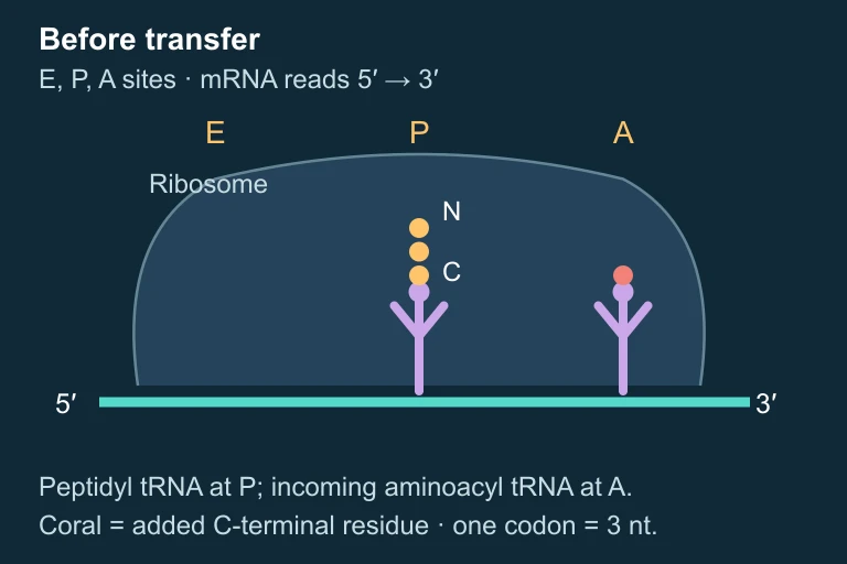 Before peptide transfer.
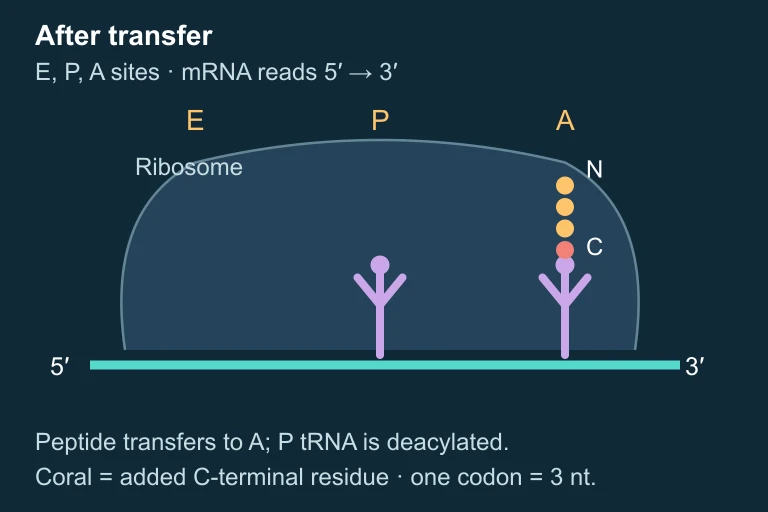 After peptide transfer.
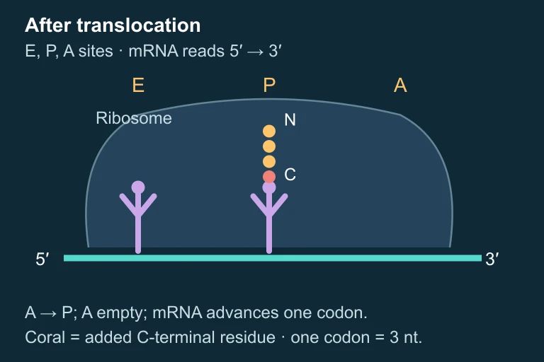 After translocation.
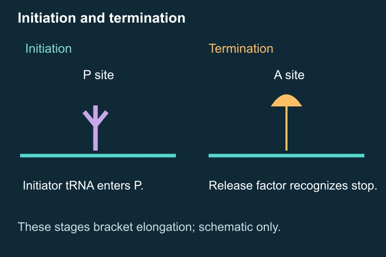 Initiation and termination.
 During elongation, a peptide of three gold residues transfers to an incoming coral amino acid in the A site, adding that residue at its C terminus. After translocation the four-residue peptidyl-tRNA occupies P; E may contain deacylated tRNA and A is empty. One codon corresponds to three nucleotides.
SECTION 5
The Genetic Code & tRNA Charging
The genetic code is the set of rules that maps each three-nucleotide codon to a specific amino acid (or a stop signal). Three properties of the code are especially important for the AP exam: it is degenerate (multiple codons can specify the same amino acid), unambiguous (each codon specifies only one amino acid), and nearly universal across all domains of life, with minor exceptions such as mitochondrial genomes and certain protists. The redundancy arises primarily at the third ("wobble") position, where non-standard base pairing is tolerated.
| Codon Feature | Detail | Significance |
|---|---|---|
| Start codon | AUG (methionine) | Sets the reading frame; always the first amino acid incorporated |
| Stop codons | UAA, UAG, UGA | Recognized by release factors, not tRNAs; signal termination |
| Wobble position | 3rd nucleotide of codon | Allows non-standard base pairing; fewer than 61 tRNAs needed |
| Degeneracy | Most amino acids: 2–6 codons | Silent mutations often occur at wobble position |
Key features of the genetic code relevant to translation
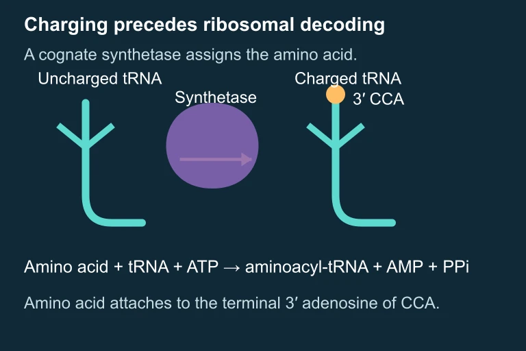 Synthetase charging and ATP consumption.
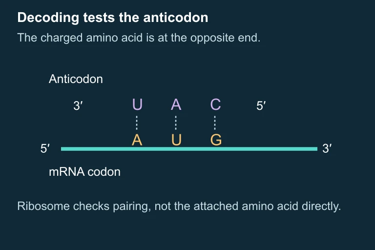 Separate AUG codon and UAC anticodon example.
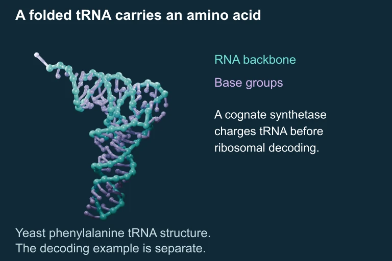 Folded yeast phenylalanine tRNA, shown as a simplified coordinate-based backbone and base representation. The AUG decoding example is a separate lesson example, not the sequence of this structure.
SECTION 6
Worked Example: From mRNA to Polypeptide
A common AP Biology question provides an mRNA sequence and asks you to determine the resulting amino acid sequence. Let's work through a complete example using the codon table.
Worked example
- 1
Step 1 — Identify the Start Codon
Given mRNA (5′→3′): GCAUAUGGUUAACUGUUGAUAA. Scan from 5′ to 3′ for the first AUG. The sequence begins GCA-UAU-GGU… but the first AUG starts at position 4: …AUG GUU AAC UGU UGA UAA. Translation begins at this AUG.Start codon identified at position 4: AUG - 2
Step 2 — Establish the Reading Frame
Starting from AUG, divide the remaining sequence into triplets: AUG | GUU | AAC | UGU | UGA | UAA. The reading frame is fixed by the start codon.Six codons identified in reading frame - 3
Step 3 — Decode Each Codon
Using the standard codon table: AUG = Met, GUU = Val, AAC = Asn, UGU = Cys, UGA = Stop. Because UGA is a stop codon, the ribosome terminates here. The downstream UAA is not translated.Amino acid sequence: Met-Val-Asn-Cys - 4
Step 4 — Consider a Mutation
If a single nucleotide deletion removes the first U in GUU, the new reading frame from AUG onward becomes: AUG | GUA | ACU | GUU | GAU | AA… The amino acid sequence changes to Met-Val-Thr-Val-Asp-… and the original stop codon is lost (frameshift). This illustrates how a frameshift mutation alters every downstream amino acid and typically eliminates proper termination.Frameshift: entirely different protein, likely nonfunctional
SECTION 7
Prokaryotic vs. Eukaryotic Translation
Although the fundamental mechanism of translation is conserved, significant differences exist between prokaryotic and eukaryotic systems. Understanding these differences is critical for AP Biology because they explain why certain antibiotics can selectively target bacterial ribosomes without harming host cells, and why gene expression regulation differs between domains.
| Feature | Prokaryotes | Eukaryotes |
|---|---|---|
| Ribosome size | 70S (30S + 50S) | 80S (40S + 60S) |
| Initiation | Shine-Dalgarno sequence; fMet-tRNA; 3 IFs | 5′ cap scanning; Met-tRNA; ≥12 eIFs |
| Coupling with transcription | Yes — co-transcriptional (no nucleus) | No — mRNA exported from nucleus first |
| Polycistronic mRNA | Common (operons) | Rare (monocistronic) |
| Post-translational processing | Limited (no ER/Golgi) | Extensive (ER, Golgi, signal peptides) |
| Antibiotic targets | Chloramphenicol, tetracycline, erythromycin | Cycloheximide (research tool only) |
Major differences in translation between prokaryotes and eukaryotes
SECTION 8
Regulation of Translation & Connections to Advanced Topics
Translation does not occur in isolation — it is subject to multiple layers of regulation that connect it to broader themes in gene expression. Cells must fine-tune protein production in response to nutrient availability, stress signals, and developmental cues. Several regulatory mechanisms are frequently tested on the AP exam.
| Regulatory Mechanism | How It Works | AP Connection |
|---|---|---|
| miRNA / siRNA | Small RNAs bind complementary mRNA sequences, triggering degradation or blocking ribosome access | Post-transcriptional regulation; RNA interference |
| 5′ cap & poly-A tail | Protect mRNA from exonucleases and promote ribosome recruitment; removal shortens mRNA lifespan | mRNA processing; stability affects protein output |
| Phosphorylation of eIF-2 | Kinases phosphorylate eIF-2 under stress, globally reducing initiation rates | Signal transduction; cellular stress response |
| Polyribosomes (polysomes) | Multiple ribosomes translate the same mRNA simultaneously, increasing protein output per transcript | Efficiency of gene expression |
| Signal peptide & SRP | N-terminal signal sequence directs ribosome to rough ER via signal recognition particle | Protein targeting; endomembrane system |
Translational regulation mechanisms and their AP Biology connections
Looking forward, understanding translation is essential for topics such as mutations and their phenotypic effects (missense, nonsense, frameshift), biotechnology applications (expressing recombinant proteins in bacterial hosts exploits universal codon usage), and evolutionary evidence (the near-universality of the genetic code strongly supports common ancestry). The AP exam frequently tests the ability to predict the effect of a point mutation on the translated protein, so practice tracing from DNA → mRNA → amino acid sequence.
SECTION 9
Practice Problems
Question 1: CONCEPTUAL
- A. Ribosomal proteins catalyze peptide bond formation between amino acids.
- B. Ribosomal RNA in the large subunit catalyzes peptide bond formation.
- C. The ribosome uses RNA as a template to synthesize more RNA molecules.
- D. The ribosome self-splices its own rRNA introns during assembly.
Show answer (B)
Question 2: BASIC
- A. 2
- B. 3
- C. 4
- D. 5
Show answer (B)
Question 3: INTERMEDIATE
- A. Aminoacyl-tRNA binding to the A site
- B. Peptide bond formation between the P-site and A-site amino acids
- C. Translocation of the ribosome along the mRNA
- D. Release factor recognition of the stop codon
Show answer (B)
Question 4: APPLIED
Show answer
Question 5: CRITICAL THINKING