
SECTION 1
Historical Context & Motivation
The discovery that genetic information flows from DNA to RNA to protein stands as one of the most consequential insights in molecular biology. After Watson and Crick elucidated the double-helical structure of DNA in 1953, the scientific community faced an urgent question: how does the information encoded in a stable, nuclear molecule direct the synthesis of proteins in the cytoplasm? The answer required identifying an intermediary molecule — messenger RNA (mRNA) — and the enzyme responsible for copying DNA into RNA, known as RNA polymerase. These discoveries launched decades of research into the mechanisms of transcription and the elaborate processing steps that convert a primary transcript into a mature, functional RNA.
1956 • Discovery of RNA Polymerase Activity
1961 • The Messenger RNA Hypothesis
1970 • Sigma Factor and Promoter Recognition
1977 • Discovery of Introns and RNA Splicing
1993 • Spliceosome Structure Emerges
The central question that unifies this lesson is: how does a cell accurately copy a specific segment of its genome into RNA, and what additional modifications must eukaryotic cells perform before that RNA can function? Understanding transcription and RNA processing is essential not only for the AP Biology exam but for appreciating how gene expression is regulated at multiple levels — from promoter access to alternative splicing — with profound implications for development, disease, and biotechnology.
SECTION 2
Core Principles of Transcription
Transcription is the process by which the information stored in a DNA template is enzymatically copied into a complementary RNA molecule. Although the fundamental logic of transcription — base pairing between DNA and incoming ribonucleotides — is conserved across all domains of life, eukaryotic transcription involves greater complexity in terms of enzyme diversity, regulatory architecture, and post-transcriptional processing. Before exploring the mechanism in detail, it is important to establish the foundational principles that govern how transcription operates.
Template-Directed Synthesis
Promoter-Driven Initiation
No Primer Required
Selective Gene Activation
Eukaryotic RNA Processing
KEY TAKEAWAY
SECTION 3
Visual Overview of Transcription
The following diagram illustrates the three major stages of transcription — initiation, elongation, and termination — showing the spatial relationship between DNA, RNA polymerase, and the growing RNA transcript. Pay particular attention to strand polarity and the direction of polymerase movement.
Note that in the diagram, the coding strand runs 5′→3′ from left to right (pink), while the template strand runs antiparallel 3′→5′ (blue). RNA polymerase reads the template strand and synthesizes the mRNA in the 5′→3′ direction, which means the mRNA sequence is complementary to the template but essentially identical to the coding strand (with U instead of T). On the AP exam, you may be asked to determine the mRNA sequence given either the template or the coding strand — always keep the base-pairing rules and strand polarity in mind.
SECTION 4
Detailed Mechanism of Transcription
Initiation: Assembling the Transcription Machinery
In eukaryotes, transcription of protein-coding genes is carried out by RNA Polymerase II (Pol II), a multi-subunit enzyme that cannot recognize promoters on its own. Instead, a series of general transcription factors (GTFs) — TFIIA, TFIIB, TFIID, TFIIE, TFIIF, and TFIIH — assemble in a stepwise manner at the core promoter to form the pre-initiation complex (PIC). The process begins when the TATA-binding protein (TBP), a subunit of TFIID, binds the TATA box approximately 25–30 base pairs upstream of the transcription start site. TFIIH then uses its helicase activity to unwind about 11–15 base pairs of DNA, creating the transcription bubble, and its kinase activity phosphorylates the C-terminal domain (CTD) of Pol II, triggering promoter clearance and the transition to elongation.
Elongation: Synthesizing the RNA Transcript
Once Pol II clears the promoter, it enters the elongation phase. The enzyme moves along the template strand in the 3′→5′ direction, adding complementary ribonucleoside triphosphates (NTPs) — ATP, UTP, GTP, and CTP — to the growing RNA chain in the 5′→3′ direction. Each incoming NTP is joined to the 3′-OH of the preceding nucleotide by a phosphodiester bond, releasing pyrophosphate (PPi), whose subsequent hydrolysis drives the reaction forward. The rate of elongation in eukaryotes is roughly 1,000–2,000 nucleotides per minute. Pol II maintains a short RNA:DNA hybrid of about 8 base pairs within the active site; behind the polymerase, the DNA double helix re-anneals and the nascent RNA strand peels away as a single-stranded molecule.
Termination: Releasing the Transcript
Termination mechanisms differ between prokaryotes and eukaryotes. In bacteria, two well-characterized pathways exist: rho-independent (intrinsic) termination, in which a GC-rich hairpin loop followed by a poly-U stretch in the nascent RNA destabilizes the RNA:DNA hybrid and causes dissociation, and rho-dependent termination, in which the rho (ρ) protein, an ATP-dependent helicase, catches up to a paused polymerase and unwinds the RNA:DNA hybrid. In eukaryotes, Pol II transcription of mRNA genes terminates via the cleavage-polyadenylation model: the pre-mRNA is cleaved at a specific site downstream of the AAUAAA polyadenylation signal, and the remaining RNA still being synthesized by Pol II is degraded by a 5′→3′ exonuclease, which eventually catches the polymerase and triggers termination.
Prokaryotes vs. Eukaryotes
SECTION 5
Eukaryotic RNA Processing
In eukaryotic cells, the primary transcript produced by RNA Polymerase II — called pre-mRNA — undergoes three critical processing events before it is exported from the nucleus as mature mRNA. These modifications occur co-transcriptionally (while transcription is still underway) and are essential for mRNA stability, proper nuclear export, and efficient translation. The three events are 5′ capping, 3′ polyadenylation, and RNA splicing.
5′ Capping
As soon as the first 20–30 nucleotides of the pre-mRNA emerge from RNA Polymerase II, capping enzymes add a 7-methylguanosine (7mG) residue to the 5′ end through an unusual 5′→5′ triphosphate linkage. This cap serves multiple functions: it protects the mRNA from degradation by 5′ exonucleases, facilitates recognition by the nuclear cap-binding complex (CBC) for export through nuclear pores, and is later recognized by the eukaryotic initiation factor eIF4E during translation initiation. Without the cap, the mRNA would be rapidly degraded and would fail to engage the ribosomal machinery.
3′ Polyadenylation
The 3′ end of the pre-mRNA is processed by endonucleolytic cleavage at a site 10–30 nucleotides downstream of the highly conserved AAUAAA polyadenylation signal. After cleavage, the enzyme poly-A polymerase (PAP) adds approximately 100–250 adenine nucleotides to form the poly-A tail. This tail enhances mRNA stability by slowing 3′ exonuclease degradation, assists in nuclear export, and stimulates translation efficiency. The length of the poly-A tail can influence mRNA lifespan — a shorter tail is associated with more rapid degradation.
RNA Splicing
Most eukaryotic protein-coding genes contain introns (non-coding intervening sequences) interspersed among exons (expressed sequences). In some human genes, introns may comprise more than 90% of the primary transcript. The removal of introns and the joining of exons is catalyzed by the spliceosome, a large ribonucleoprotein (RNP) complex composed of five small nuclear ribonucleoproteins (snRNPs: U1, U2, U4, U5, U6) and many associated proteins. Splicing proceeds through two sequential transesterification reactions. First, the 2′-OH of a conserved branch-point adenine within the intron attacks the 5′ splice site, forming a lariat intermediate. Second, the free 3′-OH of the upstream exon attacks the 3′ splice site, joining the two exons and releasing the intron lariat for degradation.
Alternative Splicing — Expanding Protein Diversity
SECTION 6
Worked Example: From DNA to Mature mRNA
Consider the following problem, which integrates transcription and RNA processing. You are given a segment of a eukaryotic gene and asked to determine the sequence of the mature mRNA.
Determining Mature mRNA Sequence
- 1
Step 1 — Identify the Given Information
A portion of a eukaryotic gene has the following coding (sense) strand sequence (written 5′→3′):5′-ATG·GCA·|·GTA·AGG·TCA·|·TTT·GCC·TAA-3′. The vertical bars (|) denote exon-intron boundaries. Exon 1 = ATG GCA, Intron = GTA AGG TCA, Exon 2 = TTT GCC TAA. The template strand is the complement read 3′→5′. - 2
Step 2 — Write the Pre-mRNA Sequence
The pre-mRNA has the same sequence as the coding strand but with U replacing T:5′-AUG·GCA·GUA·AGG·UCA·UUU·GCC·UAA-3′. This is the primary transcript before any processing.Pre-mRNA: 5′-AUG GCA GUA AGG UCA UUU GCC UAA-3′ - 3
Step 3 — Apply 5′ Capping and 3′ Polyadenylation
A 7-methylguanosine cap is added to the 5′ end (shown as 7mG–), and a poly-A tail (~100–250 As) is added to the 3′ end. The coding sequence itself is not altered by these modifications, but they are crucial structural features of the mature mRNA.7mG–5′-AUG GCA [GUA AGG UCA] UUU GCC UAA-3′-AAAAAA...A - 4
Step 4 — Remove the Intron via Splicing
The spliceosome recognizes the intron boundaries and removes the intron sequence GUA AGG UCA, joining Exon 1 directly to Exon 2. The exons are ligated by a phosphodiester bond.Mature mRNA: 7mG–5′-AUG GCA UUU GCC UAA-3′-AAAAAA...A - 5
Step 5 — Verify the Reading Frame
Reading the mature mRNA in triplet codons from the start codon AUG: AUG (Met) – GCA (Ala) – UUU (Phe) – GCC (Ala) – UAA (stop). The mRNA encodes a 4-amino-acid polypeptide: Met-Ala-Phe-Ala. Notice that the intron interrupted the coding sequence but, upon removal, the reading frame was preserved — this is a fundamental requirement of proper splicing.Polypeptide: Met-Ala-Phe-Ala
SECTION 7
Prokaryotic vs. Eukaryotic Transcription
Although the fundamental chemistry of RNA synthesis is conserved, prokaryotic and eukaryotic transcription differ significantly in terms of enzyme complexity, regulatory mechanisms, and post-transcriptional processing. The following table summarizes the key distinctions that are frequently tested on the AP Biology exam.
| Feature | Prokaryotes | Eukaryotes |
|---|---|---|
| RNA Polymerase | Single type (core enzyme + σ factor) | Three types: Pol I (rRNA), Pol II (mRNA), Pol III (tRNA, 5S rRNA) |
| Promoter Elements | −10 (Pribnow box) and −35 consensus sequences | TATA box, Inr, DPE; enhancers may be thousands of bp away |
| Transcription Factors | σ factor for initiation; few additional regulators | Multiple GTFs (TFIIA–H); Mediator complex; numerous activators/repressors |
| mRNA Processing | None (mRNA is ready for translation immediately) | 5′ cap, poly-A tail, intron splicing |
| Coupling with Translation | Yes — co-transcriptional translation | No — transcription in nucleus, translation in cytoplasm |
| Termination | Rho-dependent or rho-independent (intrinsic) | Cleavage-polyadenylation coupled with torpedo model |
| Gene Structure | Genes typically lack introns; operons group related genes | Most genes contain introns; monocistronic mRNAs |
Key differences between prokaryotic and eukaryotic transcription
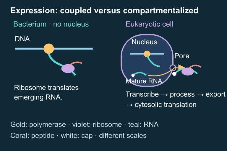 Coupled and compartmentalized expression.
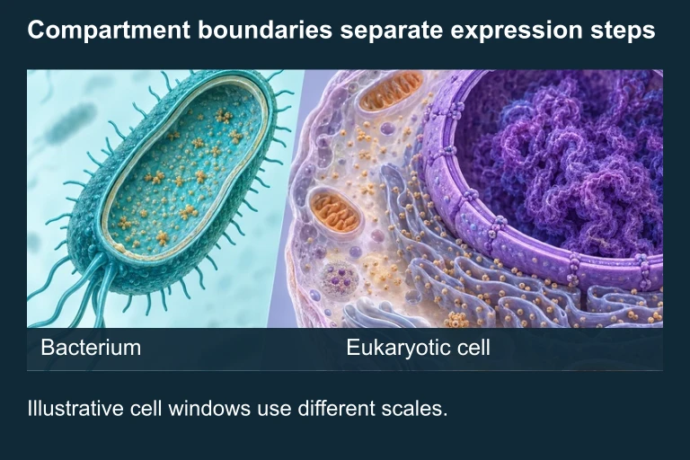 The selected bacterial model couples transcription with translation on emerging RNA. The eukaryotic comparison places transcription and processing inside a nucleus and translation after pore-mediated export to cytosol; the two cell windows use different schematic scales.
KEY TAKEAWAY
SECTION 8
Connections to Regulation and Disease
Transcription and RNA processing are not merely mechanical steps in gene expression — they are heavily regulated and serve as critical control points that determine which proteins a cell produces, when, and in what quantities. Understanding these connections is essential for the AP Biology exam and for appreciating the broader significance of gene regulation in development, cellular differentiation, and disease.
| Concept in This Lesson | Advanced Connection |
|---|---|
| Promoter recognition by Pol II | Epigenetic modifications (DNA methylation, histone acetylation) regulate promoter accessibility. CpG island hypermethylation can silence tumor suppressor genes, contributing to cancer. |
| Enhancers and transcription factors | Combinatorial control by multiple transcription factors enables cell-type-specific gene expression. Mutations in enhancers can cause developmental disorders (e.g., limb malformations from Shh enhancer mutations). |
| Alternative splicing | Aberrant splicing accounts for ~15% of human genetic diseases. For example, certain mutations in the SMN1 gene cause spinal muscular atrophy by disrupting normal splicing patterns. |
| Poly-A tail length | mRNA stability regulation through poly-A tail shortening (deadenylation) is a post-transcriptional control mechanism. MicroRNAs (miRNAs) can accelerate deadenylation and mRNA degradation. |
| 5′ cap recognition | mRNA vaccines (e.g., COVID-19) use synthetic mRNAs with modified 5′ caps and optimized UTRs to enhance stability and translation in host cells — a direct biomedical application of RNA processing principles. |
How transcription and RNA processing connect to advanced topics
As you advance beyond the foundational material covered in this lesson, keep in mind that transcription is the first — and often the most important — regulatory step in gene expression. The concepts of transcriptional regulation (operons in prokaryotes, enhancers and chromatin remodeling in eukaryotes) and post-transcriptional regulation (alternative splicing, mRNA stability, RNA interference) are deeply tested on the AP Biology exam. Future lessons will explore these regulatory layers in detail, building upon the mechanistic foundations established here.
SECTION 9
Practice Problems
Question 1: CONCEPTUAL
- A. Eukaryotic RNA polymerase makes more errors than prokaryotic RNA polymerase, requiring correction through processing.
- B. Eukaryotic genes contain introns that must be removed, and the mRNA requires modifications for stability and nuclear export, whereas prokaryotic genes generally lack introns and mRNA is translated immediately in the cytoplasm.
- C. Prokaryotic ribosomes can translate RNA with introns still present, while eukaryotic ribosomes cannot.
- D. Eukaryotic mRNA is chemically different from prokaryotic mRNA and requires capping before ribonucleotides can form phosphodiester bonds.
Show answer (B)
Question 2: BASIC
- A. 5′-AUGCCUAAG-3′
- B. 5′-ATGCCTAAG-3′
- C. 5′-GAAUCCGUA-3′
- D. 5′-UACGGAUUC-3′
Show answer (A)
Question 3: INTERMEDIATE
- A. RNA Polymerase II will stall at the mutated site and fail to complete transcription.
- B. The 5′ cap will not be added to the pre-mRNA because splicing is required before capping.
- C. The spliceosome will fail to recognize the 5′ splice site of intron 3, likely leading to intron retention or exon skipping and production of an aberrant mRNA.
- D. The poly-A tail will be added at the wrong position because splicing and polyadenylation use the same signal sequence.
Show answer (C)
Question 4: APPLIED
4 ptsShow answer
Question 5: CRITICAL THINKING
4 ptsShow answer
SUMMARY
Lesson Summary
Summary
Transcription is the process by which RNA polymerase reads the template strand of DNA (3′→5′) and synthesizes a complementary RNA molecule (5′→3′). The process proceeds through three stages: initiation at a promoter (involving general transcription factors and the pre-initiation complex in eukaryotes), elongation (NTP addition via phosphodiester bond formation), and termination (rho-dependent/independent in prokaryotes; cleavage-polyadenylation in eukaryotes).
In eukaryotes, the primary transcript (pre-mRNA) undergoes three essential processing modifications: addition of a 5′ 7-methylguanosine cap for stability and translation initiation, addition of a 3′ poly-A tail (~100–250 adenines) for stability and export, and removal of introns by the spliceosome (composed of snRNPs), which joins exons into a continuous coding sequence. Alternative splicing allows a single gene to produce multiple mRNA isoforms, vastly expanding the proteomic diversity of eukaryotic organisms. These processing steps are absent in prokaryotes, where transcription and translation are coupled in the cytoplasm.