

SECTION 1
Historical Context & Motivation
The notion that cells in a multicellular organism must communicate with one another may seem obvious today, but it took over a century of experimental work to unravel the molecular machinery underlying signal transduction. Early physiologists recognized that hormones released into the bloodstream could affect distant organs, yet the mechanism by which a chemical message at the cell surface could alter gene expression deep in the nucleus remained mysterious. The challenge was especially daunting because signaling molecules such as peptide hormones cannot cross the hydrophobic plasma membrane, demanding a relay system that bridges the extracellular and intracellular environments. Understanding this relay—from receptor activation through cascading molecular events to a cellular response—became one of the defining quests of modern cell biology.
1905 • Concept of Hormones
1957 • Discovery of Cyclic AMP
1971 • G-Protein Signaling Elucidated
1986 • Receptor Tyrosine Kinases Characterized
2012 • GPCRs and the Nobel Prize
The central question that drove this century of research can be distilled to a single puzzle: how does a water-soluble signaling molecule that cannot permeate the plasma membrane produce specific, rapid, and often amplified changes inside the cell? Signal transduction pathways provide the answer, and understanding their logic is essential for making sense of topics from embryonic development to cancer biology.
SECTION 2
Core Principles of Signal Transduction
Signal transduction pathways share a conserved three-stage architecture regardless of the specific molecules involved. First, a ligand binds a receptor during the reception stage; second, the signal is relayed and often amplified through a cascade of intracellular molecules during transduction; and third, the cell produces a response such as altered gene expression, enzyme activation, or changes in cell shape. Grasping these three stages and the molecular logic that connects them is the key to mastering signal transduction on the AP Biology exam.
Reception
Transduction
Response
Signal Amplification
Termination & Regulation
KEY TAKEAWAY
SECTION 3
Visual Overview of Signal Transduction
In the diagram above, note how the signal flows from the extracellular space through the membrane and into the cytoplasm. The receptor spans the membrane, acting as a molecular bridge. Once activated, the receptor triggers a cascade of kinases (K1 → K2 → K3), each phosphorylating and activating the next. Simultaneously, second messengers such as cAMP, Ca²⁺, and IP₃/DAG are generated; these small molecules diffuse rapidly through the cytoplasm, amplifying the signal by activating many downstream targets at once. The final outcome—changes in gene expression, metabolic enzyme activity, or cellular behavior—depends on which proteins the target cell expresses. This is why the same ligand (for example, epinephrine) can cause glycogen breakdown in liver cells, increased heart rate in cardiac cells, and smooth-muscle relaxation in bronchioles: each cell type contains a different set of downstream effectors.
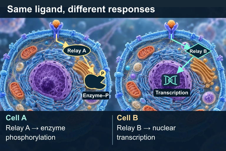 The same compatible ligand and receptor can produce different responses because the cells express different intracellular relays and effectors.
SECTION 4
Mechanisms of Transduction: Receptor Types & Cascades
G-Protein-Coupled Receptors (GPCRs)
GPCRs constitute the largest superfamily of membrane receptors in humans, with over 800 members mediating responses to hormones, neurotransmitters, and sensory stimuli. Each GPCR threads the membrane seven times (hence the alternate name seven-transmembrane receptors). On the cytoplasmic side, the receptor interacts with a heterotrimeric G protein composed of α, β, and γ subunits. In the inactive state, the Gα subunit binds GDP. Ligand binding causes the receptor to act as a guanine nucleotide exchange factor (GEF), promoting the exchange of GDP for GTP on the Gα subunit. GTP-bound Gα dissociates from the βγ dimer, and both fragments can activate downstream effectors such as adenylyl cyclase (which converts ATP to cAMP) or phospholipase C (which cleaves PIP₂ into IP₃ and DAG). The intrinsic GTPase activity of Gα eventually hydrolyzes GTP back to GDP, terminating the signal.
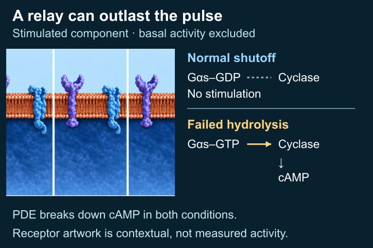 Inactive Gαs–GDP ceases to stimulate cyclase, while hydrolysis-defective Gαs–GTP can continue stimulation after the hormone pulse. PDE-mediated cAMP degradation continues in both. The selected signal-stimulated component excludes basal cyclase activity; repeated raster panels show contextual hardware, not measured states.
Receptor Tyrosine Kinases (RTKs)
RTKs are single-pass transmembrane proteins with an extracellular ligand-binding domain and an intracellular kinase domain. Ligand binding promotes dimerization—two receptor monomers come together—followed by cross-phosphorylation (autophosphorylation) of tyrosine residues on the cytoplasmic tails. These phosphotyrosines serve as docking sites for relay proteins containing SH2 domains, which activate branching downstream pathways such as the Ras–MAPK pathway (involved in cell growth and division) and the PI3K–Akt pathway (promoting cell survival). Because a single activated RTK can recruit multiple relay proteins simultaneously, RTKs are potent activators of complex, branching signal networks. Mutations that make RTKs constitutively active are common in cancers—for example, overexpression of the HER2 receptor in certain breast cancers.
Ligand-Gated Ion Channels
In the nervous system, speed of signaling is paramount. Ligand-gated ion channels are receptor proteins that open an ion-conducting pore directly upon ligand binding, bypassing the multi-step relay of GPCRs or RTKs. When acetylcholine binds the nicotinic acetylcholine receptor at a neuromuscular junction, Na⁺ ions rush into the postsynaptic cell within milliseconds, depolarizing the membrane and initiating muscle contraction. Although these channels do not involve a classical phosphorylation cascade, they exemplify the principle that extracellular signals produce intracellular changes—in this case, a change in membrane potential and ion concentration.
Intracellular Receptors
Not all signaling molecules require a membrane receptor. Hydrophobic ligands such as steroid hormones (estrogen, testosterone, cortisol) and thyroid hormones can diffuse through the plasma membrane and bind intracellular receptors located in the cytoplasm or nucleus. The ligand–receptor complex typically functions as a transcription factor, binding directly to DNA regulatory elements and altering gene expression. Because the signal bypasses the membrane-to-cytoplasm relay, these pathways generally have slower onset but longer-lasting effects compared with GPCR or RTK signaling.
SECTION 5
Signal Amplification & Second Messengers
One of the most remarkable features of signal transduction is the enormous amplification that occurs between the initial receptor activation event and the final cellular response. A single molecule of epinephrine binding a β-adrenergic receptor can activate roughly 100 G protein molecules, each of which activates an adenylyl cyclase molecule that synthesizes approximately 100 cAMP molecules, and each cAMP-activated protein kinase A (PKA) can phosphorylate around 10 substrates. Through this multiplicative cascade, a single ligand–receptor interaction can mobilize millions of product molecules—a phenomenon central to the fight-or-flight response.
Key Second Messengers
| Second Messenger | Produced By | Primary Target(s) | Cellular Effects |
|---|---|---|---|
| cAMP | Adenylyl cyclase (from ATP) | Protein kinase A (PKA) | Glycogen breakdown, gene regulation, ion channel modulation |
| Ca²⁺ | Released from ER via IP₃-gated channels | Calmodulin, PKC | Muscle contraction, exocytosis, apoptosis |
| IP₃ | Phospholipase C (from PIP₂) | IP₃-gated Ca²⁺ channels on ER | Ca²⁺ release from ER stores |
| DAG | Phospholipase C (from PIP₂) | Protein kinase C (PKC) | Cell growth, differentiation |
Major second messengers in signal transduction pathways
AP EXAM TIP
SECTION 6
Worked Example: Tracing the Epinephrine Signaling Pathway
The following worked example traces the epinephrine (adrenaline) signaling pathway in a liver cell from ligand binding to glycogen breakdown. This is one of the most commonly tested signal transduction pathways on the AP Biology exam.
Epinephrine → Glycogen Breakdown in Liver Cells
- 1
Step 1 — Reception
Epinephrine, released by the adrenal medulla during the fight-or-flight response, circulates through the blood and binds to a β-adrenergic receptor (a GPCR) on the surface of a liver cell. The binding is highly specific, driven by complementary shape and non-covalent interactions between the ligand and the receptor's extracellular domain.Receptor undergoes conformational change - 2
Step 2 — G-Protein Activation
The conformational change on the cytoplasmic face of the receptor causes it to act as a GEF for the associated Gα subunit. GDP is exchanged for GTP on Gα, which dissociates from Gβγ. The activated Gα-GTP subunit diffuses along the membrane to encounter adenylyl cyclase.Gα-GTP activates adenylyl cyclase - 3
Step 3 — Second Messenger Production
Activated adenylyl cyclase catalyzes the conversion of ATP to cyclic AMP (cAMP). Each adenylyl cyclase molecule produces many cAMP molecules per second, providing the first major amplification step. cAMP diffuses through the cytoplasm and binds the regulatory subunits of protein kinase A (PKA), releasing its catalytic subunits.PKA catalytic subunits are activated - 4
Step 4 — Phosphorylation Cascade
Active PKA phosphorylates phosphorylase kinase, which in turn phosphorylates and activates glycogen phosphorylase. Each kinase in the cascade activates many substrate molecules, further amplifying the signal.Glycogen phosphorylase is activated - 5
Step 5 — Cellular Response
Activated glycogen phosphorylase catalyzes the cleavage of glucose monomers from glycogen, releasing glucose-1-phosphate into the cytoplasm. The glucose is subsequently exported from the liver cell into the bloodstream, raising blood sugar to fuel muscles and the brain during the stress response.Glycogen → glucose released into blood - 6
Step 6 — Signal Termination
The signal is terminated at multiple levels: the Gα subunit hydrolyzes GTP → GDP (returning to its inactive state), phosphodiesterase degrades cAMP to AMP, and protein phosphatases remove phosphate groups from the kinase cascade enzymes. Without active termination, the pathway would remain constitutively on.Pathway returns to baseline; signal is off
SECTION 7
Comparing Major Receptor Types
Different receptor types offer different trade-offs in speed, amplification, and specificity. The table below summarizes the key features of each major receptor class, helping you distinguish among them for exam questions that present an unfamiliar signaling scenario and ask you to identify the receptor type involved.
| Feature | GPCR | RTK | Ligand-Gated Ion Channel | Intracellular Receptor |
|---|---|---|---|---|
| Membrane spans | 7 transmembrane helices | 1 transmembrane helix (dimerizes) | Multi-subunit pore | Not membrane-bound |
| Ligand type | Hormones, neurotransmitters, odorants | Growth factors (EGF, PDGF, insulin) | Neurotransmitters (ACh, GABA) | Steroid/thyroid hormones, NO |
| Speed | Seconds to minutes | Minutes | Milliseconds | Hours (gene expression) |
| Amplification | High (second messengers) | High (branching cascades) | Low (direct ion flow) | Low (direct gene regulation) |
| Key mechanism | G protein → second messenger | Dimerization → cross-phosphorylation | Ion channel opens → ion flux | Ligand–receptor acts as transcription factor |
| Example | β-adrenergic receptor (epinephrine) | EGF receptor, HER2 | Nicotinic ACh receptor | Estrogen receptor |
Comparison of the four major receptor classes in cell signaling
KEY TAKEAWAY
SECTION 8
Signal Transduction & Disease: Connections to Cancer and Pharmacology
Disruptions in signal transduction pathways are at the heart of many human diseases, most notably cancer. The AP Biology curriculum explicitly connects cell signaling to the cell cycle and its regulation, so understanding how mutations in signaling components lead to uncontrolled proliferation is both clinically important and exam-relevant.
| Normal Signaling Component | When Mutated / Dysregulated | Disease Connection |
|---|---|---|
| Ras (G protein in RTK pathway) | Constitutively active (GTPase activity lost); stuck in GTP-bound "on" state | ~30% of all human cancers harbor Ras mutations (lung, pancreatic, colorectal) |
| HER2 (RTK) | Gene amplification → receptor overexpression → excessive growth signals | HER2-positive breast cancer; treated with monoclonal antibody trastuzumab (Herceptin) |
| p53 (transcription factor) | Loss-of-function mutation → cell cannot halt cell cycle or trigger apoptosis | Mutated in >50% of cancers; known as "guardian of the genome" |
| Cholera toxin (exogenous) | Modifies Gα subunit to prevent GTP hydrolysis → cAMP levels remain permanently high | Severe diarrhea in cholera: Cl⁻ and H₂O secretion into intestinal lumen is unregulated |
How disrupted signaling leads to disease
These examples highlight a unifying theme: signal transduction pathways are tightly regulated systems, and any mutation that locks a pathway in the "on" state (an oncogene) or disables a pathway that restrains growth (a tumor suppressor) can contribute to cancer. Modern pharmacology targets these pathways: drugs like imatinib (Gleevec) inhibit a constitutively active tyrosine kinase in chronic myelogenous leukemia, while caffeine inhibits phosphodiesterase to prolong cAMP signaling. Recognizing these connections between normal signaling and pathology will prepare you for both the AP exam and any future coursework in molecular biology or medicine.
LOOKING AHEAD
SECTION 9
Practice Problems
Question 1: CONCEPTUAL
- A. Intracellular steroid receptor
- B. G-protein-coupled receptor (GPCR)
- C. Ligand-gated ion channel
- D. Receptor tyrosine kinase (RTK)
Show answer (B)
Question 2: BASIC CALCULATION
- A. 10
- B. 100
- C. 1,000
- D. 10,000
Show answer (C)
Question 3: INTERMEDIATE
- A. The cell will be unable to respond to any growth factors.
- B. The cell will undergo continuous cell division even without growth factor stimulation.
- C. The cell will immediately undergo apoptosis.
- D. Cyclic AMP levels will be permanently elevated in the cell.
Show answer (B)
Question 4: APPLIED
4 ptsShow answer
Question 5: CRITICAL THINKING
4 ptsShow answer
SUMMARY
Lesson Summary
Summary
Signal transduction pathways convert extracellular signals into intracellular responses through three conserved stages: reception (ligand binds receptor), transduction (relay and amplification via phosphorylation cascades and second messengers such as cAMP, Ca²⁺, IP₃, and DAG), and response (altered gene expression, enzyme activity, or cell behavior). The four major receptor classes—GPCRs, receptor tyrosine kinases, ligand-gated ion channels, and intracellular receptors—differ in speed, amplification potential, and downstream targets, allowing cells to tailor their responses to diverse signals.
Signal amplification is a hallmark of transduction cascades, with multiplicative gains at each enzymatic step enabling a single ligand to generate millions of product molecules. Equally important is signal termination, achieved through phosphatases, GTPase activity, and second-messenger degradation by enzymes like phosphodiesterase. Disruptions in these pathways—such as constitutively active Ras oncogenes or loss-of-function mutations in tumor suppressors like p53—are central to cancer biology. For the AP exam, remember that the same ligand can produce different responses in different cell types because the response depends on the cell's unique complement of intracellular proteins, not solely on the identity of the signal.