
SECTION 1
Historical Context & Motivation
The study of proteins has been central to biology since the term was coined in the nineteenth century. Dutch chemist Gerardus Johannes Mulder first described these nitrogen-rich substances in 1838, and Swedish chemist Jöns Jacob Berzelius suggested naming them after the Greek word proteios, meaning "of first importance." That label proved prescient: proteins turned out to be the primary molecular workhorses of life, executing functions from catalysis and transport to immune defense and signal transduction.
1838 • Naming of Proteins
1902 • The Peptide Bond Hypothesis
1951 • α-Helix and β-Sheet Described
1958 • First Protein Structure Solved
1972 • Anfinsen's Dogma
A fundamental question links all of these milestones: how does the linear sequence of amino acids in a polypeptide give rise to the precise three-dimensional shape that determines a protein's biological function? This relationship between structure and function is the organizing theme of protein biology and a core idea tested throughout the AP Biology curriculum.
SECTION 2
Core Principles & Definitions
Proteins are polymers assembled from amino acid monomers. Twenty different amino acids are encoded by the genetic code, each sharing a common backbone—an amino group (−NH₂), a carboxyl group (−COOH), a hydrogen atom, and an α-carbon—but differing in their R group (side chain). The chemical properties of R groups—nonpolar, polar, acidic, or basic—determine how a polypeptide folds and how the mature protein interacts with substrates, ligands, and other macromolecules.
Amino Acid Monomers
Peptide Bond Formation
Four Levels of Structure
Structure Determines Function
SECTION 3
Amino Acid Structure & the Peptide Bond
The diagram above highlights two essential ideas. First, all twenty amino acids share the same backbone architecture—only the R group varies, and that variation is sufficient to produce the full range of protein diversity observed in living systems. Second, the peptide bond is formed through a condensation (dehydration synthesis) reaction in which the carboxyl group of one amino acid reacts with the amino group of the next, releasing one water molecule per bond. This reaction is catalyzed by the ribosome during translation and is reversed during hydrolysis, the addition of water to break covalent bonds.
SECTION 4
The Four Levels of Protein Structure
Protein organization is described at four hierarchical levels, each stabilized by specific types of chemical interactions. Understanding these levels is essential because structure dictates function—the loss of any structural level can render a protein nonfunctional.
Primary Structure
The primary structure is the unique linear sequence of amino acids in a polypeptide chain, read from the N-terminus (free amino group) to the C-terminus (free carboxyl group). Because the sequence is encoded by the gene, even a single nucleotide mutation can substitute one amino acid for another and alter the protein's shape and activity—sickle-cell disease results from a single valine-for-glutamic-acid substitution at position 6 of β-globin.
Secondary Structure
Local regions of the polypeptide fold into repeating patterns stabilized by hydrogen bonds between backbone N−H and C=O groups. The two most common motifs are the α-helix, a right-handed coil with hydrogen bonds every 3.6 residues, and the β-pleated sheet, formed by hydrogen bonds between adjacent polypeptide strands running parallel or antiparallel to each other.
Tertiary Structure
The overall three-dimensional shape of a single polypeptide chain constitutes its tertiary structure. It arises from interactions among R groups, including hydrophobic interactions (nonpolar R groups clustering in the interior), ionic bonds (between charged R groups), hydrogen bonds (between polar R groups), van der Waals forces, and disulfide bridges (covalent S−S bonds between cysteine residues). In aqueous environments, hydrophobic interactions are often the dominant driving force for folding.
Quaternary Structure
When a functional protein consists of two or more polypeptide subunits, their spatial arrangement constitutes the quaternary structure. Hemoglobin, for instance, is a tetramer of two α-globin and two β-globin subunits. The same types of non-covalent interactions that stabilize tertiary structure also hold subunits together. Not all proteins possess quaternary structure—myoglobin, for example, functions as a single polypeptide.
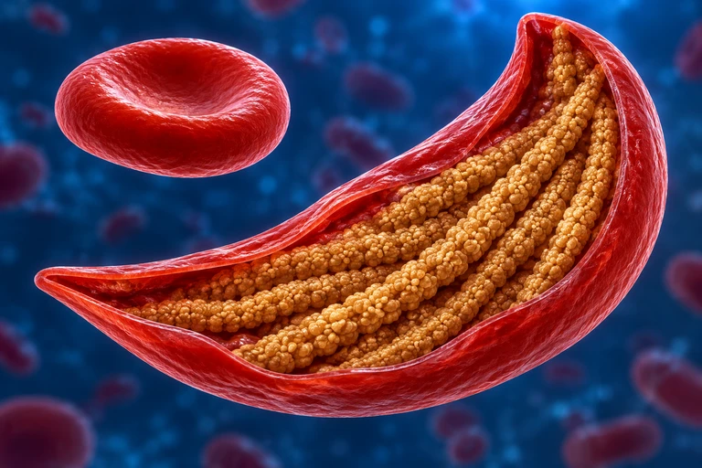 The mature β-globin Glu6→Val6 substitution replaces a charged surface residue with a nonpolar one. Under low oxygen, HbS can assemble into fibers that deform red cells.
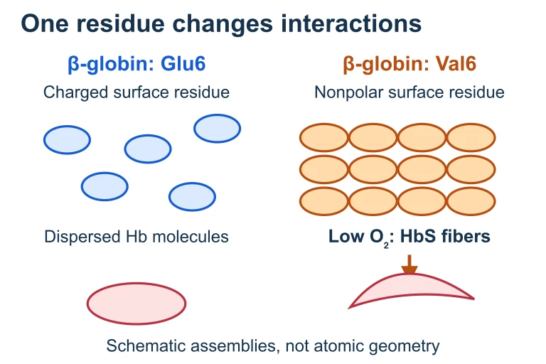 The mature β-globin Glu6→Val6 substitution replaces a charged surface residue with a nonpolar one. Under low oxygen, HbS can assemble into fibers that deform red cells.
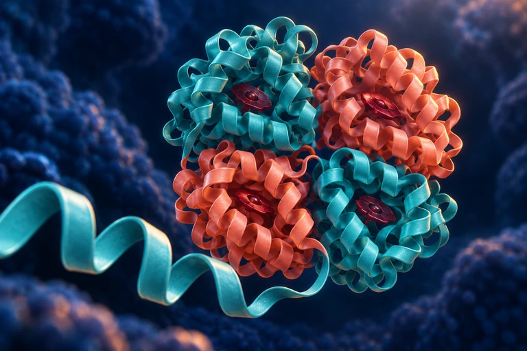 Protein organization spans amino-acid sequence, local secondary structure, a folded chain and multi-chain assemblies. An α-helix uses backbone hydrogen bonds from residue i to i+4; hemoglobin contains two α and two β chains.
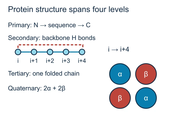 Protein organization spans amino-acid sequence, local secondary structure, a folded chain and multi-chain assemblies. An α-helix uses backbone hydrogen bonds from residue i to i+4; hemoglobin contains two α and two β chains.
SECTION 5
Protein Diversity & Functional Classes
A remarkably small alphabet of twenty amino acids generates an essentially infinite variety of proteins by varying sequence length and composition. Even a short polypeptide of 100 residues has 20¹⁰⁰ possible sequences—a number that dwarfs the estimated atoms in the observable universe. Cells exploit this diversity to produce proteins that fall into several broad functional categories.
| Class | Example | Function |
|---|---|---|
| Enzyme | DNA polymerase | Catalyzes DNA replication |
| Structural | Collagen | Provides tensile strength in connective tissue |
| Transport | Hemoglobin | Carries O₂ in red blood cells |
| Defensive | Immunoglobulin (antibody) | Binds and neutralizes antigens |
| Signaling | Insulin | Peptide hormone regulating blood glucose |
| Motor | Myosin | Generates force for muscle contraction |
| Storage | Casein | Stores amino acids in milk |
Functional classes of proteins with representative examples
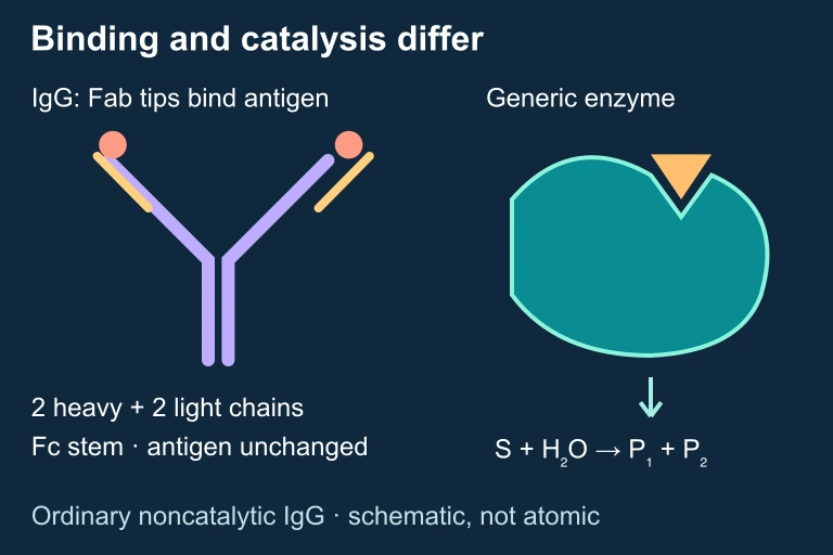 IgG binding and enzyme catalysis
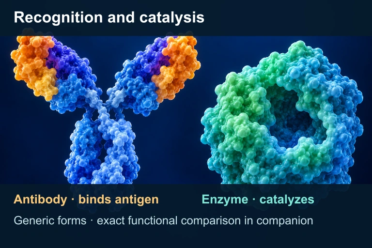 An ordinary noncatalytic IgG recognizes antigen at its Fab tips without changing it; a generic enzyme catalyzes a reaction. The selected schematic has two heavy and two light chains; protein forms are illustrative, not atomic structures. Catalytic antibodies exist and are outside this example.
SECTION 6
Worked Example — Analyzing Protein Structure & Denaturation
The following example walks through a scenario commonly tested on the AP Biology exam: predicting how environmental changes affect protein structure and function.
Worked example
- 1
Step 1 — Identify the Protein and Its Normal Environment
Consider the enzyme lactase, which catalyzes the hydrolysis of lactose into glucose and galactose. Lactase functions optimally at approximately 37 °C (human body temperature) and neutral pH. - 2
Step 2 — Apply the Environmental Change
If the temperature is raised to 80 °C, the increased kinetic energy causes rapid molecular vibrations that disrupt the weak non-covalent interactions (hydrogen bonds, ionic bonds, hydrophobic interactions) stabilizing the enzyme's tertiary structure. - 3
Step 3 — Predict the Structural Consequence
The loss of non-covalent interactions causes the polypeptide to unfold, a process called denaturation. Note that the primary structure (peptide bonds) remains intact; only the higher-order structure is disrupted.Tertiary (and secondary) structure is lost → active site shape changes - 4
Step 4 — Predict the Functional Consequence
Because lactase's catalytic activity depends on the precise geometry of its active site, denaturation prevents the substrate (lactose) from binding effectively. The reaction rate drops to near zero, even though the substrate concentration remains the same.Enzymatic activity is abolished due to active site deformation - 5
Step 5 — Consider Reversibility
Some proteins can refold when returned to favorable conditions (as Anfinsen demonstrated with ribonuclease), but many proteins, especially large multi-domain enzymes, aggregate irreversibly when denatured at high temperatures—similar to cooking an egg, where the albumin proteins cannot return to their native state.
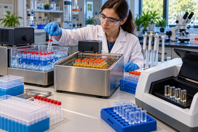 To distinguish prior temperature exposure from immediate reaction conditions, return all enzyme treatments to the same assay temperature and pH. Recovery after cooling differs from persistent activity loss after heating.
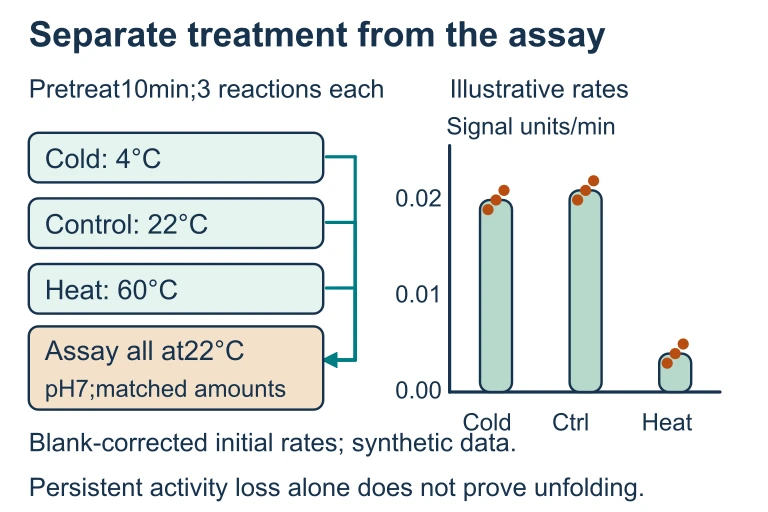 To distinguish prior temperature exposure from immediate reaction conditions, return all enzyme treatments to the same assay temperature and pH. Recovery after cooling differs from persistent activity loss after heating.
SECTION 7
Comparing Bonds & Interactions in Protein Structure
A common source of confusion on the AP exam is conflating the different types of bonds and interactions that contribute to each structural level. The table below clarifies which interactions operate at which level and their relative strengths.
| Interaction | Type | Structural Level(s) | Relative Strength |
|---|---|---|---|
| Peptide bond | Covalent | Primary | Very strong |
| Hydrogen bond (backbone) | Non-covalent | Secondary | Weak individually; strong collectively |
| Hydrophobic interaction | Non-covalent | Tertiary, Quaternary | Major folding driver in aqueous solutions |
| Ionic bond (salt bridge) | Non-covalent | Tertiary, Quaternary | Moderate; pH-sensitive |
| Disulfide bridge | Covalent (S−S) | Tertiary | Strong; resists denaturation |
| Van der Waals forces | Non-covalent | Tertiary, Quaternary | Weakest individually; additive |
Summary of stabilizing interactions across the four levels of protein structure
SECTION 8
Proteins in the Broader Biological Context
Protein biology does not exist in isolation; it connects directly to genetics, evolution, and cellular regulation. The central dogma of molecular biology—DNA → RNA → Protein—places proteins as the ultimate functional products of gene expression. Mutations in DNA alter the primary structure of proteins, which may change higher-order structure, and natural selection acts on the phenotypic consequences of those structural changes.
| Concept in This Lesson | Advanced Connection |
|---|---|
| Primary structure determined by gene | Gene regulation (operons, transcription factors) controls which proteins are made and when |
| Enzyme specificity (active site shape) | Enzyme kinetics, allosteric regulation, competitive & noncompetitive inhibition |
| Denaturation by pH/temperature | Chaperone proteins (Hsp70, GroEL) assist folding; misfolding diseases (Alzheimer's, prion diseases) |
| Quaternary structure (hemoglobin) | Cooperative binding, allosteric effects, and the oxygen-hemoglobin dissociation curve |
| Amino acid R-group diversity | Post-translational modifications (phosphorylation, glycosylation) expand functional diversity |
Connections between foundational protein concepts and advanced AP Biology topics
As you progress through the AP Biology curriculum, you will encounter proteins at every turn—membrane channel proteins in cell communication, receptor proteins in signal transduction, and motor proteins in cell division. Mastering the structural principles covered here provides the conceptual scaffolding for understanding each of these advanced topics.
SECTION 9
Practice Problems
Question 1: CONCEPTUAL
- A. Secondary structure
- B. Primary structure
- C. Tertiary structure
- D. Quaternary structure
Show answer (B)
Question 2: BASIC
- A. 150
- B. 151
- C. 149
- D. 148
Show answer (C)
Question 3: INTERMEDIATE
- A. The enzyme's primary structure is disrupted, breaking peptide bonds.
- B. The enzyme's tertiary structure is destabilized, likely reducing catalytic activity.
- C. The enzyme gains a new catalytic function due to the added charge.
- D. The enzyme's secondary structure is eliminated because α-helices require nonpolar residues.
Show answer (B)
Question 4: APPLIED
Show answer
Question 5: CRITICAL THINKING