
SECTION 1
Historical Context & Motivation
The question of how cells maintain their internal environment while remaining open to nutrients, signals, and waste removal has driven biological inquiry for over a century. Early microscopists recognized that cells possess a boundary, but the molecular architecture of that boundary—the plasma membrane—remained elusive until converging discoveries in chemistry, physics, and electron microscopy revealed its lipid bilayer organization. Understanding how this membrane was conceptualized over time provides essential context for appreciating the fluid mosaic model that guides modern cell biology.
1895 • Overton's Lipid Hypothesis
1925 • Gorter & Grendel Bilayer Evidence
1935 • Davson–Danielli Sandwich Model
1972 • Singer–Nicolson Fluid Mosaic Model
2000s • Lipid Rafts & Membrane Microdomains
The central question that connects all of these milestones is deceptively simple: how does a structure just 7–8 nanometers thick simultaneously serve as a physical barrier, a communication hub, and a selectively permeable gateway? Answering this question requires understanding the chemistry of phospholipids, the behavior of membrane proteins, and the thermodynamic principles that give the bilayer its remarkable properties.
SECTION 2
Core Principles & Definitions
The plasma membrane is far more than a passive container; it is a dynamic, amphipathic assembly whose properties emerge from the interplay between its lipid, protein, and carbohydrate components. Several foundational principles underpin its architecture and function, each contributing to the membrane's capacity for selective permeability, cell recognition, and signal transduction.
Amphipathic Phospholipids
Fluid Mosaic Model
Selective Permeability
Membrane Asymmetry
Cholesterol as Fluidity Buffer
KEY TAKEAWAY
SECTION 3
Visual Explanation — The Fluid Mosaic
The following diagram illustrates a cross-sectional view of the plasma membrane according to the fluid mosaic model. Pay close attention to the orientation of phospholipids, the positioning of integral versus peripheral proteins, and the location of cholesterol molecules within the bilayer.
Several features of this diagram merit close attention. First, notice the asymmetry: carbohydrate chains extend only from the extracellular face, while peripheral proteins are shown on the cytoplasmic face. Second, cholesterol is intercalated between phospholipid tails, not sitting on the membrane surface; this positioning allows it to interact with fatty acid chains and modulate their packing. Third, the integral protein spans both leaflets, exposing domains to both the extracellular fluid and the cytoplasm—a feature essential for receptor and transport functions. Finally, the channel protein creates a hydrophilic pore through the hydrophobic core, enabling ions and small polar molecules to traverse the membrane without contacting the lipid tails directly.
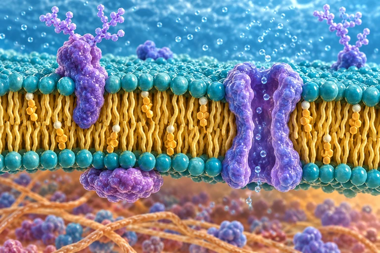 Polar phospholipid heads face water on both sides of the membrane while hydrophobic tails face inward. Carbohydrate chains on glycoproteins extend from the extracellular face.
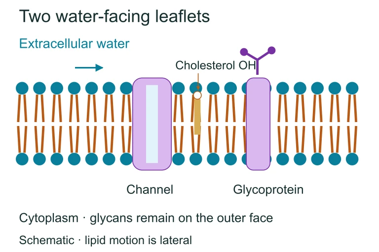 Polar phospholipid heads face water on both sides of the membrane while hydrophobic tails face inward. Carbohydrate chains on glycoproteins extend from the extracellular face.
SECTION 4
Transport Mechanisms Across the Membrane
Because the plasma membrane is selectively permeable, cells rely on multiple transport mechanisms to move substances across the bilayer. These mechanisms fall into two broad categories—passive transport, which requires no cellular energy input, and active transport, which requires ATP hydrolysis or coupling to another energy source. The thermodynamic driving force for passive transport is the free energy change associated with moving a solute down its concentration gradient, while active transport moves solutes against their gradient.
FREE ENERGY OF SOLUTE TRANSPORT
Where R = gas constant (8.314 J·mol⁻¹·K⁻¹), T = temperature in Kelvin, [S]ᵢₙ and [S]ₒᵤₜ = solute concentrations inside and outside the cell, z = net charge of the solute, F = Faraday's constant (96,485 C·mol⁻¹), and ΔΨ = membrane potential. For uncharged solutes, the electrical term (zFΔΨ) drops out, and transport depends solely on the concentration ratio.
When ΔG is negative, movement of the solute across the membrane is thermodynamically favorable and can proceed passively. When ΔG is positive, the cell must couple the transport event to an energy-releasing process—typically ATP hydrolysis—to drive the solute uphill. This distinction underpins every transport modality discussed in AP Biology.
OSMOLARITY AND WATER POTENTIAL
Water potential (Ψ) equals the sum of solute potential (Ψₛ, always ≤ 0 for solutions) and pressure potential (Ψₚ). Water moves from regions of higher Ψ to lower Ψ by osmosis. For animal cells lacking cell walls, Ψₚ is typically 0, making Ψₛ the dominant determinant of water movement.
AP Exam Connection
| Transport Type | Energy Source | Direction | Examples |
|---|---|---|---|
| Simple Diffusion | None (ΔG < 0) | Down gradient | O₂, CO₂, steroid hormones |
| Facilitated Diffusion | None (ΔG < 0) | Down gradient via protein | Glucose (GLUT1), ions (K⁺ channels), aquaporins (water) |
| Osmosis | None (ΔG < 0) | High Ψ → Low Ψ | Water across membrane |
| Primary Active Transport | ATP hydrolysis | Against gradient | Na⁺/K⁺-ATPase, H⁺ pump, Ca²⁺-ATPase |
| Secondary Active (Cotransport) | Ion gradient (indirect ATP) | Against gradient (coupled) | Na⁺/glucose symporter, Na⁺/H⁺ antiporter |
| Endocytosis / Exocytosis | ATP (vesicle formation) | Bulk movement | Phagocytosis, pinocytosis, receptor-mediated endocytosis, secretion |
Summary of major transport mechanisms across the plasma membrane
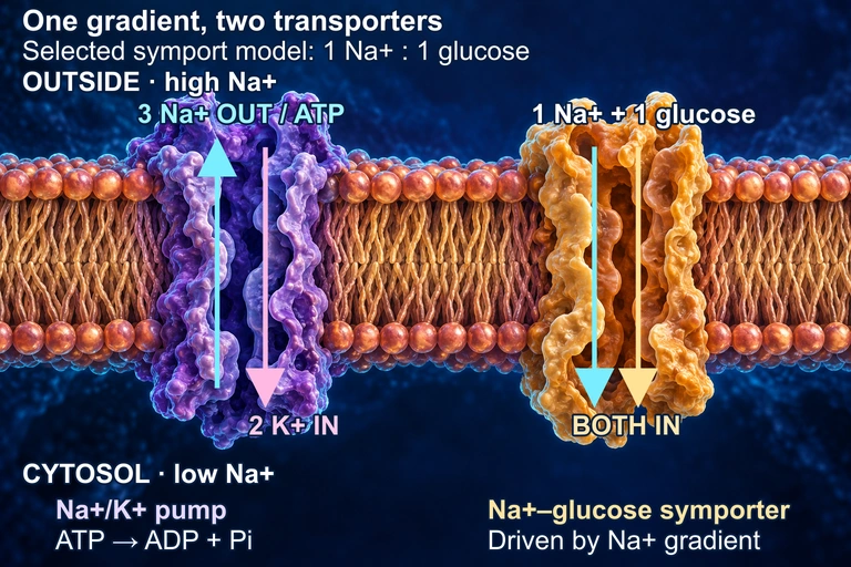 In this conceptual membrane, the Na+/K+ ATPase uses one ATP to move three Na+ out and two K+ in, maintaining a sodium electrochemical gradient. A separate symporter uses that gradient to bring Na+ and glucose inward; the 1:1 ratio is a selected teaching model and varies among transporters. The cutaways do not represent constitutively open pores: both transport cycles and the protein shapes are schematic. The juxtaposition does not assert a shared membrane face in a particular tissue.
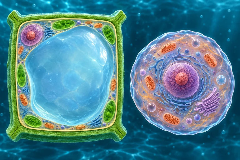 For nonpenetrating solutes, a plant wall limits expansion as water enters. Water loss retracts the protoplast from the wall during plasmolysis; animal cells lack this rigid wall.
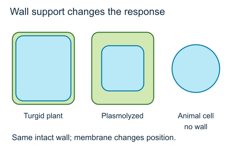 For nonpenetrating solutes, a plant wall limits expansion as water enters. Water loss retracts the protoplast from the wall during plasmolysis; animal cells lack this rigid wall.
SECTION 5
Membrane Protein Classification & Function
Proteins constitute approximately 50% of membrane mass and are responsible for virtually all of the membrane's specific functions—from catalysis and transport to cell signaling and structural support. Understanding the functional classes of membrane proteins is critical for AP Biology, as exam questions frequently ask students to connect protein structure to cellular processes.
Each of these protein functions can be tested on the AP exam in the context of a larger biological process. For instance, signal transduction connects membrane receptors to Unit 4 (Cell Communication), while cell-cell recognition ties into the immune system's ability to distinguish self from non-self. Transport proteins recur throughout discussions of neuronal signaling (Na⁺/K⁺-ATPase), kidney function, and photosynthesis (proton pumps in thylakoid membranes). Recognizing these connections enables you to apply membrane protein knowledge across multiple AP Biology units.
SECTION 6
Worked Example — Predicting Osmotic Behavior
A classic AP Biology problem asks you to predict what happens to a cell placed in solutions of varying tonicity. Let's work through a scenario that integrates water potential, osmolarity, and the selective permeability of the plasma membrane.
Red Blood Cell in Three Solutions
- 1
Step 1 — Define the Problem
A human red blood cell (RBC) with an internal solute concentration of 0.9% NaCl is placed into three beakers: (A) 0.9% NaCl, (B) 0.1% NaCl, and (C) 5.0% NaCl. Predict the direction of net water movement and the resulting cell morphology in each case. Assume the membrane is permeable to water (via aquaporins) but impermeable to NaCl. - 2
Step 2 — Classify Tonicity
Tonicity describes the relative concentration of non-penetrating solutes outside versus inside the cell. Beaker A is isotonic (0.9% = 0.9%). Beaker B is hypotonic (0.1% < 0.9%). Beaker C is hypertonic (5.0% > 0.9%).A = isotonic; B = hypotonic; C = hypertonic - 3
Step 3 — Predict Water Movement Using Water Potential
For animal cells, Ψₚ ≈ 0, so Ψ = Ψₛ. Higher solute concentration → more negative Ψₛ → lower Ψ. Water moves from high Ψ to low Ψ. In Beaker A, Ψ is equal on both sides → no net movement. In Beaker B, the external solution has higher Ψ (less negative Ψₛ) → water enters the cell. In Beaker C, the external solution has lower Ψ (more negative Ψₛ) → water exits the cell.A: no net flow; B: water enters RBC; C: water exits RBC - 4
Step 4 — Predict Cell Morphology
In Beaker A, the RBC maintains its normal biconcave shape. In Beaker B, the influx of water causes the cell to swell; if the hypotonic stress is severe, the cell undergoes hemolysis (bursting), since RBCs lack a cell wall to resist turgor pressure. In Beaker C, water loss causes the cell to shrink and develop a spiky, irregular shape—a process called crenation.A: normal shape; B: hemolysis (lysis); C: crenation - 5
Step 5 — Compare with a Plant Cell
A plant cell in a hypotonic environment absorbs water but does not lyse because the rigid cell wall exerts an opposing pressure potential (Ψₚ > 0). Instead, the cell becomes turgid, which is the ideal state for plant cells. In a hypertonic environment, the plasma membrane pulls away from the cell wall—a phenomenon called plasmolysis. This contrast between animal and plant cell responses is a high-yield topic on the AP exam.
SECTION 7
Passive vs. Active Transport — Strengths & Limitations
Cells exploit both passive and active mechanisms, and each has distinct advantages and constraints. The table below highlights key comparisons that help contextualize why cells require multiple transport modalities rather than relying on diffusion alone.
| Feature | Passive Transport | Active Transport |
|---|---|---|
| Energy Requirement | None—driven by concentration/electrochemical gradient | ATP or coupled ion gradient |
| Direction | Down the gradient only | Against the gradient |
| Saturability | Simple diffusion: not saturable. Facilitated: saturable (limited by number of transport proteins) | Saturable—limited by number of pump proteins and ATP availability |
| Specificity | Simple diffusion: low (depends on size/polarity). Facilitated: high (protein-dependent) | High—pump proteins are substrate-specific |
| Biological Role | Equilibration of small molecules; gas exchange; osmosis | Maintaining ion gradients, nutrient uptake against gradients, signal generation |
| Limitation | Cannot establish or maintain concentration gradients; ceases at equilibrium | Energetically costly; disrupted by metabolic poisons (e.g., cyanide) |
Comparison of passive and active transport mechanisms
KEY TAKEAWAY
SECTION 8
Connection to Advanced Topics
The plasma membrane is not an isolated topic; it is the physical foundation for processes examined throughout the AP Biology curriculum and beyond. Understanding membrane dynamics opens doors to cell signaling (Unit 4), cellular energetics (Unit 3), and even evolutionary biology (Unit 7). The table below contrasts foundational membrane concepts with their more advanced extensions.
| AP Biology Foundation | Advanced Extension |
|---|---|
| Fluid mosaic model: lipids and proteins move laterally | Lipid rafts and membrane microdomains organize signaling complexes; FRAP experiments measure diffusion coefficients of membrane components |
| Selective permeability via transport proteins | Patch-clamp electrophysiology measures single ion channel conductance; channelopathies (e.g., cystic fibrosis) result from defective membrane proteins |
| Receptor-ligand binding initiates signal transduction | G-protein coupled receptors (GPCRs) represent ~34% of FDA-approved drug targets; receptor tyrosine kinases (RTKs) drive oncogenic signaling in cancer |
| Endosymbiotic origin of mitochondria/chloroplasts (double membranes) | Inner mitochondrial membrane hosts the electron transport chain; proton gradient across this membrane drives chemiosmotic ATP synthesis |
| Glycoprotein markers on cell surfaces | MHC class I and II molecules present antigen peptides; CAR-T cell therapy engineers T cells to recognize specific tumor surface antigens |
Connecting AP Biology membrane concepts to advanced biomedical topics
For students continuing into college-level biochemistry or cell biology, the membrane emerges as a central organizing theme. The chemiosmotic model, for instance, depends entirely on the impermeability of the inner mitochondrial membrane to protons—a property rooted in the same phospholipid bilayer principles covered in this lesson. Similarly, understanding viral entry mechanisms (e.g., SARS-CoV-2 spike protein binding to ACE2 receptors) requires knowledge of membrane protein structure and endocytosis. Mastering the plasma membrane at the AP level provides the conceptual scaffolding for these more advanced investigations.
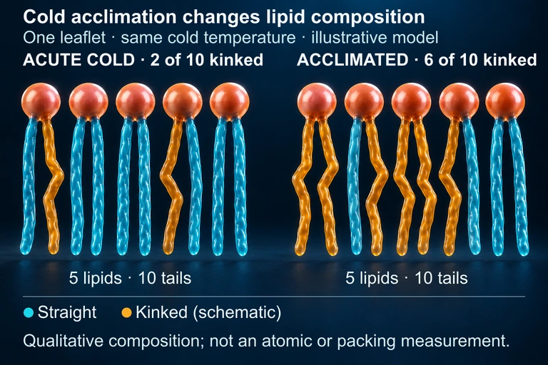 Some organisms acclimate to cold by increasing unsaturated fatty-acid content, helping maintain membrane fluidity. This qualitative composition model compares leaflets at the same cold temperature; it is not an atomic structure or packing measurement.
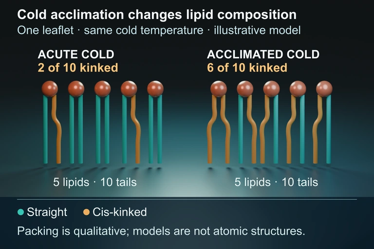 Authored geometry companion
SECTION 9
Practice Problems
Question 1: CONCEPTUAL
- A. Passive diffusion of oxygen across the membrane
- B. Cell-cell recognition and immune identification
- C. Active transport of sodium and potassium ions
- D. Maintenance of membrane fluidity at low temperatures
Show answer (B)
Question 2: BASIC CALCULATION
- A. Ψ = −0.5 MPa; water moves into the cell
- B. Ψ = −1.1 MPa; water moves out of the cell
- C. Ψ = −0.5 MPa; water moves out of the cell
- D. Ψ = +0.3 MPa; no net water movement
Show answer (A)
Question 3: INTERMEDIATE
- A. Simple diffusion; the lipid bilayer becomes saturated with glucose molecules
- B. Facilitated diffusion via carrier proteins; the plateau occurs because all carrier proteins are occupied (saturated)
- C. Primary active transport via a glucose pump; the plateau occurs because ATP supply is exhausted
- D. Endocytosis; the plateau occurs because the membrane surface area limits vesicle formation
Show answer (B)
Question 4: APPLIED
4 ptsShow answer
Question 5: CRITICAL THINKING
4 ptsShow answer
SUMMARY
Plasma Membrane — Summary
Summary
The plasma membrane is a phospholipid bilayer organized according to the fluid mosaic model, with integral proteins spanning the bilayer and peripheral proteins associated with its surfaces. Cholesterol buffers membrane fluidity across temperature ranges, while the glycocalyx of carbohydrate chains on the extracellular face mediates cell-cell recognition and immune identity. The membrane's selective permeability allows small nonpolar molecules to diffuse freely while requiring transport proteins for ions and large polar solutes.
Transport across the membrane occurs via passive mechanisms (simple diffusion, facilitated diffusion, osmosis) that require no energy input, or via active transport (pumps, cotransporters, endocytosis/exocytosis) powered by ATP hydrolysis or coupled ion gradients. Water potential (Ψ = Ψₛ + Ψₚ) governs osmotic water movement, with animal cells susceptible to hemolysis in hypotonic solutions and crenation in hypertonic ones. Plant cells resist lysis through turgor pressure but undergo plasmolysis in hypertonic environments. These membrane principles connect broadly to cell signaling, energetics, and immune function across the AP Biology curriculum.