
SECTION 1
Historical Context & Motivation
One of the most fundamental distinctions in biology separates prokaryotic cells—which lack membrane-bound organelles—from eukaryotic cells, which possess elaborate internal compartments such as the nucleus, mitochondria, endoplasmic reticulum, and Golgi apparatus. Understanding how and why eukaryotic cells evolved this internal complexity is central to modern cell biology and evolutionary theory. The question of how a relatively simple ancestral cell gave rise to the highly organized, compartmentalized eukaryotic cell represents one of the great puzzles in the history of life on Earth, and its resolution draws on evidence from microscopy, molecular phylogenetics, biochemistry, and genomics.
1883 • Andreas Schimper's Chloroplast Hypothesis
1905 • Merezhkowsky Proposes Symbiogenesis
1967 • Lynn Margulis and Endosymbiotic Theory
1970s–1990s • Molecular and Genomic Confirmation
2010s–Present • Asgard Archaea and Eukaryogenesis
The central question this lesson addresses is: How did the elaborate internal membrane systems and organelles of eukaryotic cells originate from simpler ancestral cells? The answer involves two intertwined processes—endosymbiosis and the invagination and elaboration of internal membranes—that together produced the compartmentalized architecture essential for eukaryotic complexity.
SECTION 2
Core Principles of Cell Compartmentalization
Cell compartmentalization refers to the partitioning of the cytoplasm into distinct membrane-bound regions, each maintaining a unique chemical environment optimized for specific metabolic functions. This structural organization allows eukaryotic cells to run incompatible biochemical reactions simultaneously—for example, the acidic hydrolytic environment of the lysosome operates mere micrometers away from the near-neutral pH of the cytoplasm. Several core principles underlie the origin and maintenance of compartmentalization, and understanding them is essential for grasping why eukaryotic cells function so differently from their prokaryotic counterparts.
Endosymbiotic Origin
Endomembrane System Evolution
Selective Permeability & Transport
Metabolic Efficiency
Gene Transfer to the Nucleus
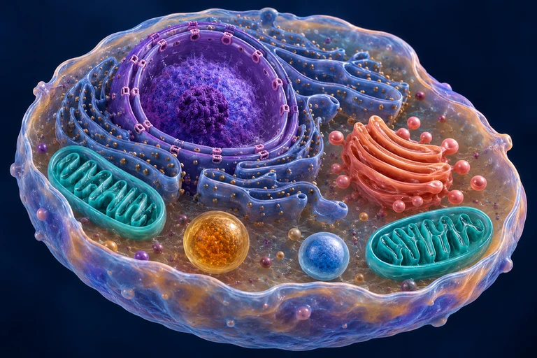 Membranes separate cytosol from organelle lumens and maintain distinct chemical environments. The nuclear outer membrane connects with the endoplasmic reticulum; mitochondrial and peroxisomal protein import follows routes distinct from ER–Golgi vesicle traffic.
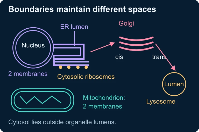 Membranes separate cytosol from organelle lumens and maintain distinct chemical environments. The nuclear outer membrane connects with the endoplasmic reticulum; mitochondrial and peroxisomal protein import follows routes distinct from ER–Golgi vesicle traffic.
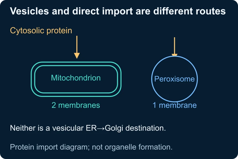 Mitochondrial and peroxisomal proteins can enter from the cytosol through direct-import routes distinct from ER–Golgi vesicle transport.
SECTION 3
Visual Explanation: Endosymbiosis & Membrane Invagination
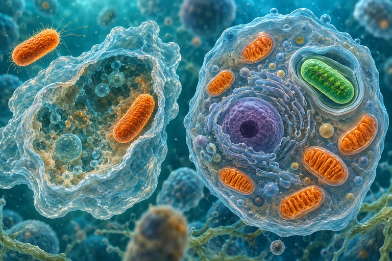
The visual above captures the two complementary processes that together gave rise to the compartmentalized eukaryotic cell. The endomembrane system (including the nuclear envelope, ER, and Golgi) likely arose through progressive invagination of the ancestral plasma membrane, a process that would have been favored because it increased the surface area available for membrane-bound reactions and allowed the cell to segregate its genetic material from potentially damaging cytoplasmic reactions. In contrast, mitochondria and chloroplasts arose through endosymbiotic events in which the host cell engulfed—but did not digest—a smaller prokaryote. Their two bounding membranes form part of the evidence for their evolutionary history. Molecular relationships and organelle genomes provide more direct tests of bacterial ancestry than assigning each membrane to a presumed engulfment step.
SECTION 4
Mechanisms of Endosymbiosis & Endomembrane Evolution
Primary Endosymbiosis: Mitochondria
The most widely supported model holds that the mitochondrial endosymbiosis occurred approximately 1.5 to 2 billion years ago, when an archaeal host cell—likely related to the Asgard archaea—engulfed an alphaproteobacterium. The engulfed bacterium was already capable of aerobic respiration, a metabolic advantage in an increasingly oxygen-rich atmosphere. Over time, a mutualistic relationship developed: the host supplied organic substrates, and the endosymbiont provided far more ATP through oxidative phosphorylation than the host could generate by fermentation alone. This metabolic payoff—roughly 15 to 18 times more ATP per glucose molecule—likely drove strong selective pressure for retaining the endosymbiont.
Primary Endosymbiosis: Chloroplasts
A second endosymbiotic event occurred when an early eukaryote (one that already possessed mitochondria) engulfed a photosynthetic cyanobacterium. This event gave rise to the chloroplast lineage and is the basis for all photosynthetic eukaryotes, including plants, green algae, and red algae. The chloroplast retains its own circular DNA and 70S ribosomes, replicates by binary fission, and possesses a double membrane—all hallmarks of its endosymbiotic origin. In some lineages, secondary endosymbiosis occurred when a non-photosynthetic eukaryote engulfed a photosynthetic eukaryote, producing plastids with three or four membranes, as seen in brown algae and euglenids.
Endomembrane System: The Invagination Model
The origin of the nuclear envelope and associated endomembrane system is explained by the invagination hypothesis. According to this model, infoldings of the plasma membrane of an ancestral prokaryote-like cell progressively extended inward, eventually surrounding the cell's DNA and forming a double-membraned nucleus. These same infoldings also gave rise to the endoplasmic reticulum (ER), which remains physically continuous with the outer nuclear membrane in modern cells—a structural relic of this shared origin. The Golgi apparatus, lysosomes, and vesicles subsequently differentiated from this primitive endomembrane network, likely through specialization of membrane composition and protein sorting machinery.
Endosymbiotic Gene Transfer (EGT)
A critical step in the integration of endosymbionts was the massive transfer of genes from the endosymbiont's genome to the host's nuclear genome—a process called endosymbiotic gene transfer (EGT). Modern mitochondrial genomes encode only about 37 genes (in humans), compared to the roughly 1,500 genes whose protein products function within the mitochondrion. The vast majority of mitochondrial proteins are encoded by nuclear genes, synthesized on cytoplasmic ribosomes, and imported into mitochondria via transit peptides recognized by the TOM/TIM translocase complexes. This dependence on the host genome for most of its proteins has rendered the mitochondrion incapable of independent life—an evolutionary point of no return that cements the obligate relationship.
 Mitochondrial ancestry traces to alphaproteobacteria. Primary plastid ancestry traces to cyanobacteria acquired by a eukaryotic lineage that already had mitochondria.
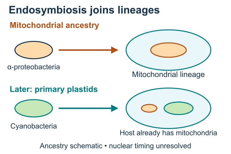 Mitochondrial ancestry traces to alphaproteobacteria. Primary plastid ancestry traces to cyanobacteria acquired by a eukaryotic lineage that already had mitochondria.
SECTION 5
Evidence for Endosymbiotic Origin
The endosymbiotic theory is one of the most strongly supported hypotheses in biology, resting on multiple independent lines of evidence. For the AP Biology exam, you should be able to articulate each line of evidence and explain how it supports the model that mitochondria originated from alphaproteobacteria and chloroplasts from cyanobacteria.
The convergence of these independent lines of evidence makes the endosymbiotic origin of mitochondria and chloroplasts among the best-supported evolutionary hypotheses. The circular DNA, 70S ribosomes, and binary fission of these organelles are all bacterial features retained from the original endosymbiont. Phylogenetic analyses of ribosomal RNA sequences place mitochondria firmly within the alphaproteobacterial clade and chloroplasts within the cyanobacterial clade, establishing not just endosymbiotic origin but identifying the specific prokaryotic lineages involved. Furthermore, the inner membranes of these organelles contain cardiolipin (in mitochondria) and bacterial-type lipids—lipid compositions consistent with their bacterial ancestry rather than with the host cell's plasma membrane.
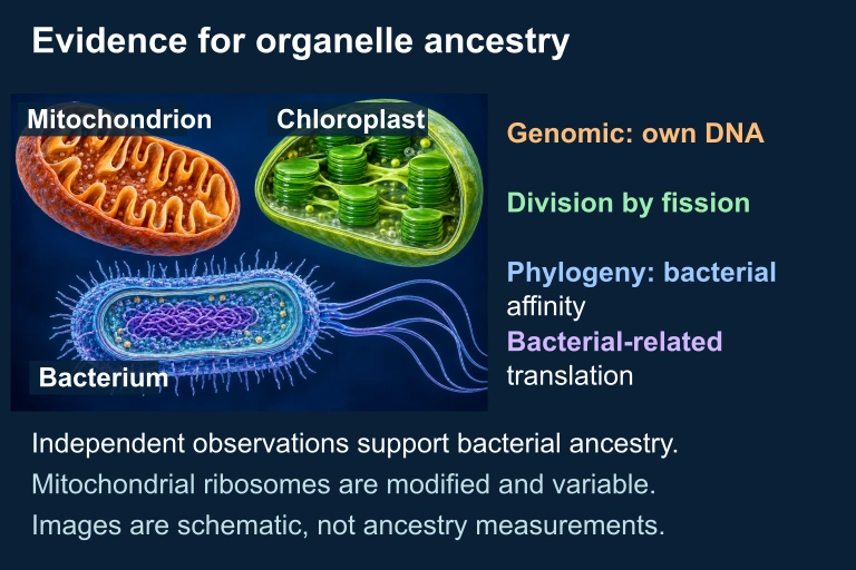 Independent evidence from organelle genomes, division, phylogeny and bacterial-related translation machinery supports bacterial ancestry of mitochondria and chloroplasts. Mitochondrial ribosomes are evolutionarily modified and variable. The organism illustrations are schematic and do not establish measurements or universal genome topology.
SECTION 6
Worked Example: Analyzing Evidence for Endosymbiosis
A common AP Biology task requires you to evaluate evidence and construct an argument supporting the endosymbiotic origin of organelles. Let us walk through a structured approach to this type of analysis.
Worked example
- 1
Step 1 — Identify the Claim
Mitochondria originated from a free-living alphaproteobacterium that was engulfed by an ancestral archaeal host cell approximately 1.5–2 billion years ago. This claim must be supported by multiple lines of evidence.Claim: Mitochondria are descended from endosymbiotic alphaproteobacteria. - 2
Step 2 — Cite Structural Evidence
Mitochondria possess a double membrane, consistent with the expected product of phagocytosis. The inner membrane, which houses the electron transport chain, has a lipid composition (including cardiolipin) that closely resembles bacterial plasma membranes rather than the eukaryotic endomembrane system. Mitochondria also contain their own circular DNA molecules, which lack histones—mirroring the genomic organization of free-living bacteria.Double membrane + circular DNA + bacterial lipids = structural evidence - 3
Step 3 — Cite Molecular/Phylogenetic Evidence
Mitochondrial ribosomal RNA (rRNA) sequences, when compared to databases of prokaryotic 16S rRNA, cluster phylogenetically with alphaproteobacteria such as Rickettsia and Rhodospirillum. Additionally, mitochondria contain 70S ribosomes (the bacterial type), not the 80S ribosomes found in the eukaryotic cytoplasm. This ribosome size difference is functionally significant—antibiotics that target 70S ribosomes (such as chloramphenicol) inhibit mitochondrial protein synthesis but do not affect cytoplasmic translation.rRNA phylogeny + 70S ribosomes + antibiotic sensitivity = molecular evidence - 4
Step 4 — Cite Reproductive Evidence
Mitochondria reproduce by binary fission, the same mechanism used by free-living bacteria. They cannot be synthesized de novo by the cell; new mitochondria arise only from pre-existing mitochondria. This semi-autonomous replication is consistent with the behavior expected of an organism that was once independently reproducing.Binary fission + inability to form de novo = reproductive evidence - 5
Step 5 — Synthesize the Argument
The structural, molecular, phylogenetic, and reproductive evidence converge on the same conclusion: mitochondria share too many features with free-living alphaproteobacteria for these similarities to have arisen by convergent evolution. The most parsimonious explanation is that mitochondria are derived endosymbionts whose genomes have been largely transferred to the host nucleus, leaving them obligately dependent on the host cell.Conclusion: Multiple independent lines of evidence strongly support the endosymbiotic origin of mitochondria.
SECTION 7
Comparing Prokaryotic and Eukaryotic Organization
Understanding the advantages and trade-offs of compartmentalization requires contrasting prokaryotic and eukaryotic cellular organization. While prokaryotes are enormously successful and occupy virtually every habitat on Earth, eukaryotic compartmentalization enabled qualitatively new levels of cellular complexity, including larger cell size, multicellularity, and sophisticated gene regulation.
| Feature | Prokaryotic Cell | Eukaryotic Cell |
|---|---|---|
| Internal membranes | Absent (some have simple infoldings, e.g., thylakoids in cyanobacteria) | Extensive endomembrane system: ER, Golgi, lysosomes, vesicles |
| DNA organization | Circular, in nucleoid region; no nuclear envelope | Linear chromosomes enclosed in double-membrane nucleus; histones |
| Cell size | Typically 1–10 µm | Typically 10–100 µm; compartmentalization supports larger volume |
| Metabolic separation | Reactions occur in cytoplasm or on plasma membrane; limited segregation | Opposing pathways compartmentalized (e.g., oxidation in mitochondria, reduction in cytoplasm) |
| Gene regulation | Transcription and translation coupled in cytoplasm | Transcription in nucleus; mRNA processing; translation in cytoplasm—allows post-transcriptional regulation |
| Energy organelles | Plasma membrane houses ETC components | Mitochondria (and chloroplasts in photosynthetic cells) with dedicated membranes |
Structural and functional comparison of prokaryotic and eukaryotic cellular organization
SECTION 8
Connections to Advanced Topics & Modern Research
The endosymbiotic origin of organelles connects to several broader themes tested on the AP Biology exam and pursued in active research. Understanding these connections deepens your ability to reason across units and apply compartmentalization concepts in novel contexts.
| AP Biology Concept | Connection to Compartmentalization |
|---|---|
| Cellular respiration (Unit 3) | The mitochondrial matrix and inner membrane are specialized compartments for the citric acid cycle and oxidative phosphorylation, respectively. The proton gradient across the inner membrane depends on compartmentalization to function. |
| Photosynthesis (Unit 3) | Chloroplast thylakoid membranes compartmentalize the light reactions, while the Calvin cycle occurs in the stroma. The thylakoid lumen accumulates H⁺ ions for chemiosmosis—a direct consequence of membrane compartmentalization. |
| Evolution (Unit 7) | Endosymbiosis is a major mechanism of evolutionary change that does not fit neatly into gradualist models. It demonstrates that symbiotic mergers can drive rapid, large-scale evolutionary transitions. |
| Gene expression (Unit 6) | The nuclear envelope separates transcription from translation, enabling RNA processing (splicing, capping, polyadenylation) and providing additional regulatory checkpoints absent in prokaryotes. |
| Maternal inheritance | Mitochondrial DNA is inherited maternally in most organisms because the egg contributes nearly all cytoplasm. This non-Mendelian inheritance pattern is a direct consequence of endosymbiotic origin and organelle semi-autonomy. |
Cross-unit connections to compartmentalization
Current research on Asgard archaea is reshaping our understanding of eukaryogenesis. These archaea, discovered in deep-sea sediments near Loki's Castle hydrothermal vents, possess homologs of eukaryotic genes involved in membrane remodeling, vesicle trafficking, and cytoskeletal dynamics. The emerging model suggests that the archaeal host already had some capacity for membrane manipulation before the mitochondrial endosymbiosis event, providing the cellular machinery needed to engulf and retain the alphaproteobacterial endosymbiont. Whether the nucleus formed before, during, or after mitochondrial acquisition remains an area of active debate, and future genomic and structural studies of Asgard archaea may finally resolve this question.
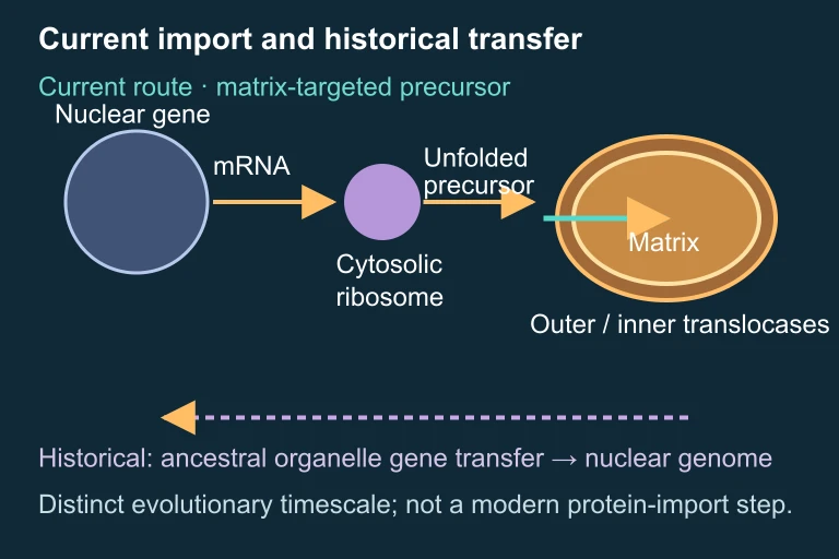 Current import and historical transfer.
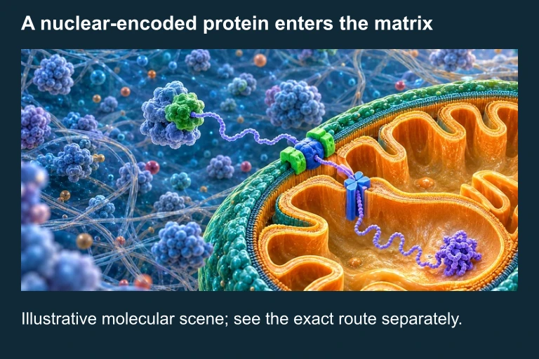 Many mitochondrial proteins are encoded in the nucleus, translated in the cytosol and imported into the organelle. Illustration is schematic where scale or molecular structure is simplified.
SECTION 9
Practice Problems
Question 1: CONCEPTUAL
- A. Mitochondria are enclosed by a double membrane.
- B. Mitochondria contain 70S ribosomes and circular DNA whose sequences are phylogenetically related to alphaproteobacteria.
- C. Mitochondria carry out oxidative phosphorylation.
- D. Mitochondria are present in nearly all eukaryotic cells.
Show answer (B)
Question 2: BASIC
- A. Synthesis of histones on cytoplasmic ribosomes
- B. Transcription of nuclear genes encoding glycolytic enzymes
- C. Translation of mitochondrial-encoded subunits of the electron transport chain
- D. Replication of nuclear DNA during S phase
Show answer (C)
Question 3: INTERMEDIATE
- A. The plastid arose from a single primary endosymbiosis followed by two additional rounds of membrane invagination.
- B. A non-photosynthetic eukaryote engulfed a photosynthetic eukaryote that already contained a double-membraned chloroplast, adding two additional membranes through the engulfment.
- C. Four independent endosymbiotic events each contributed one membrane layer to the plastid.
- D. The plastid replicated its membranes as a result of gene duplication events affecting membrane biosynthesis genes.
Show answer (B)
Question 4: APPLIED
4 ptsShow answer
Question 5: CRITICAL THINKING
4 pts