
SECTION 1
Historical Context & Motivation
The concept of heritable change has fascinated biologists since the rediscovery of Mendelian genetics at the turn of the twentieth century, but the molecular basis for such change remained elusive for decades. Early geneticists observed that organisms sometimes produced offspring with novel, stable traits—features that could not be explained by simple recombination of existing alleles. Hugo de Vries coined the term mutation in 1901 to describe these sudden, heritable variations in the evening primrose Oenothera lamarckiana, although many of the "mutations" he documented were later attributed to chromosomal rearrangements. Despite this imprecision, the term endured because it captured a genuine phenomenon: the spontaneous generation of new genetic variation.
Understanding mutations gained practical urgency during the mid-twentieth century when researchers linked radiation exposure to increased mutation rates, and later when the molecular revolution revealed that mutations are alterations in the nucleotide sequence of DNA. Today, mutation sits at the intersection of molecular biology, medicine, and evolutionary theory—it is the ultimate source of all genetic variation upon which natural selection acts, and it is the molecular basis of genetic diseases ranging from sickle cell anemia to many cancers.
1901 • De Vries and the Mutation Theory
1927 • Muller's X-ray Experiments
1953 • Watson & Crick Structure of DNA
1966 • The Genetic Code Cracked
2003 • Human Genome Project Completed
The central question this lesson addresses is deceptively simple: how do changes in DNA sequence arise, what are their molecular consequences, and why do some mutations profoundly alter phenotype while others are phenotypically silent? Answering this question requires integrating knowledge of DNA replication fidelity, the degeneracy of the genetic code, protein structure–function relationships, and the regulatory architecture of the genome.
SECTION 2
Core Principles & Definitions
A mutation is any permanent change in the nucleotide sequence of an organism's DNA (or RNA, in certain viruses). Mutations range from single-nucleotide alterations to massive chromosomal rearrangements, and their biological significance depends on context—where in the genome they occur, whether they affect coding or regulatory sequences, and whether they change the amino acid sequence of a protein product. The following core principles organize the study of mutations in AP Biology.
Mutations Are Random
Scale Matters
Germ-Line vs. Somatic
Phenotypic Effect Varies
Mutation Is the Raw Material of Evolution
KEY TAKEAWAY
SECTION 3
Visual Explanation — Point Mutation Types
The diagram below illustrates the four major categories of point mutations—silent, missense, nonsense, and frameshift—and their effects on the mRNA codon sequence and resulting polypeptide. Each column shows how a single change in the DNA template strand propagates through transcription and translation. Pay special attention to the frameshift column, where an insertion or deletion shifts the entire reading frame downstream of the mutation, typically producing a nonfunctional protein.
Several key observations emerge from this diagram. First, silent mutations exploit the degeneracy of the genetic code—because multiple codons encode the same amino acid, a nucleotide substitution in the third (wobble) position of a codon frequently produces a synonymous codon. Second, missense mutations substitute one amino acid for another, and their phenotypic impact depends heavily on whether the substitution is chemically conservative (e.g., leucine → isoleucine) or radical (e.g., glycine → glutamate). Third, nonsense mutations introduce a premature stop codon (UAA, UAG, or UGA), producing a truncated polypeptide that is usually nonfunctional and targeted for degradation. Finally, frameshift mutations caused by insertions or deletions of nucleotides that are not multiples of three are typically the most devastating because they corrupt every codon downstream of the alteration.
SECTION 4
Mechanisms of Mutagenesis
Mutations can arise through several distinct molecular mechanisms, and the AP Biology curriculum groups them into two broad categories: spontaneous mutations that result from normal cellular processes and induced mutations caused by environmental agents called mutagens. Understanding these mechanisms is essential because the AP exam frequently asks students to predict mutation outcomes based on the type of mutagenic event.
Spontaneous Mutations
During DNA replication, DNA polymerase occasionally incorporates the wrong nucleotide. The intrinsic error rate of replication (before proofreading) is approximately 10⁻⁵ errors per base pair. The 3ʹ→5ʹ proofreading exonuclease activity of the polymerase corrects most of these errors, reducing the rate to about 10⁻⁷. Post-replicative mismatch repair (MMR) further reduces the rate to approximately 10⁻⁹ to 10⁻¹⁰ per base pair per replication. Other spontaneous events include tautomeric shifts (rare hydrogen-bonding isomers of bases that mispair), depurination (loss of a purine base creating an abasic site), and deamination (conversion of cytosine to uracil, which pairs with adenine instead of guanine, causing a C·G → T·A transition after the next round of replication).
Induced Mutations
External agents significantly increase mutation rates above the spontaneous background. These mutagens fall into three categories: chemical mutagens (e.g., base analogs like 5-bromouracil, alkylating agents like ethyl methanesulfonate, and intercalating agents like ethidium bromide), physical mutagens (ultraviolet light causing thymine dimers, ionizing radiation causing double-strand breaks), and biological mutagens (transposable elements, or transposons, that insert into new genomic locations and disrupt gene function). Intercalating agents deserve special note because they insert between adjacent base pairs, distorting the helix and causing the replication machinery to insert or skip a nucleotide—resulting in frameshift mutations.
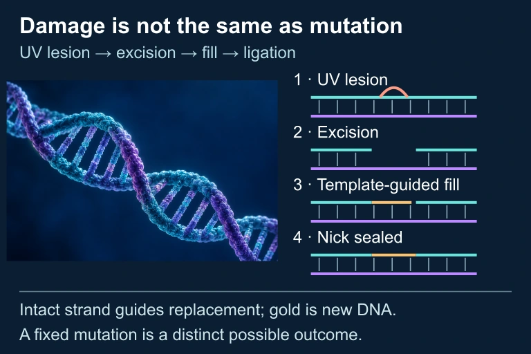 A UV-induced lesion can be excised; an intact complementary strand guides gap filling, and ligase seals the remaining nick. DNA damage and a fixed sequence mutation are distinct events. The helix is illustrative and does not depict a measured structure or specific lesion.
AP EXAM TIP
SECTION 5
Detailed Classification of Mutations
Mutations can be classified along several independent axes: by scale (point vs. chromosomal), by molecular mechanism (substitution vs. insertion/deletion), and by effect on the gene product. For the AP Biology exam, the most commonly tested classification is by phenotypic effect on the encoded protein. The table below provides a comprehensive reference for the major mutation types you are expected to recognize, along with the molecular basis and a classic biological example for each.
| Mutation Type | Molecular Change | Effect on Protein | Classic Example |
|---|---|---|---|
| Silent (synonymous) | Nucleotide substitution; new codon specifies the same amino acid | No change in amino acid sequence | CUU → CUC (both encode leucine) |
| Missense | Nucleotide substitution; new codon specifies a different amino acid | Single amino acid change; effect ranges from benign to lethal | Sickle cell disease: GAG → GUG (Glu → Val in β-globin at position 6) |
| Nonsense | Nucleotide substitution creates a premature stop codon (UAA, UAG, UGA) | Truncated, usually nonfunctional protein | Some forms of β-thalassemia; Duchenne muscular dystrophy |
| Frameshift (insertion) | One or more nucleotides inserted (not multiples of 3) | Reading frame shifts; all downstream amino acids altered; often produces premature stop | Tay-Sachs disease (4-bp insertion in HEXA gene) |
| Frameshift (deletion) | One or more nucleotides deleted (not multiples of 3) | Same as insertion frameshift | Cystic fibrosis (ΔF508 is a 3-bp deletion—an in-frame deletion, not a frameshift; however, single-bp deletions in CFTR also cause CF) |
| In-frame indel | Insertion or deletion of exactly 3 (or multiple of 3) nucleotides | Adds or removes amino acid(s) without shifting the reading frame | ΔF508 in cystic fibrosis (loss of Phe at position 508 of CFTR protein) |
Classification of point mutations by phenotypic effect on the encoded protein
Chromosomal-Scale Mutations
Beyond point mutations, large-scale chromosomal rearrangements also constitute mutations and appear on the AP exam. Deletions remove entire segments of a chromosome. Duplications produce extra copies of a chromosomal segment, which can serve as raw material for the evolution of new gene functions through subsequent divergence. Inversions reverse the orientation of a segment within a chromosome, potentially disrupting genes at the breakpoints or altering gene regulation by repositioning enhancers or silencers. Translocations move a segment from one chromosome to another; the Philadelphia chromosome (a reciprocal translocation between chromosomes 9 and 22) creates the BCR-ABL fusion oncogene that drives chronic myelogenous leukemia. Finally, aneuploidy (gain or loss of individual chromosomes, such as trisomy 21 in Down syndrome) and polyploidy (entire extra sets of chromosomes, common in plants) represent changes in chromosome number rather than structure.
SECTION 6
Worked Example — Predicting Mutation Consequences
Consider the following scenario: a segment of the template (antisense) strand of a gene reads 3ʹ–TAC GGA CTC AGA ATT–5ʹ. A single nucleotide substitution changes the ninth nucleotide (C in CTC) to A. Determine the wild-type and mutant mRNA sequences, identify the amino acids encoded, classify the mutation, and predict its phenotypic consequence.
Single Nucleotide Substitution Analysis
- 1
Step 1 — Write the wild-type mRNA
The mRNA is synthesized 5ʹ→3ʹ as the complement of the template strand (read 3ʹ→5ʹ). Template: 3ʹ–TAC GGA CTC AGA ATT–5ʹ. mRNA: 5ʹ–AUG CCU GAG UCU UAA–3ʹ. Remember, in RNA, uracil replaces thymine.Wild-type mRNA: 5ʹ–AUG CCU GAG UCU UAA–3ʹ - 2
Step 2 — Translate the wild-type mRNA
Using the codon table: AUG = Met (start), CCU = Pro, GAG = Glu, UCU = Ser, UAA = Stop.Wild-type polypeptide: Met–Pro–Glu–Ser (STOP) - 3
Step 3 — Introduce the mutation and write the mutant mRNA
The ninth nucleotide (C in the third codon CTC) is changed to A, making the template codon ATC. Template: 3ʹ–TAC GGA ATC AGA ATT–5ʹ. Mutant mRNA: 5ʹ–AUG CCU UAG UCU UAA–3ʹ.Mutant mRNA: 5ʹ–AUG CCU UAG UCU UAA–3ʹ - 4
Step 4 — Translate the mutant mRNA
AUG = Met, CCU = Pro, UAG = STOP. Translation terminates prematurely at the third codon. The ribosome never reaches the Ser codon or the original stop codon.Mutant polypeptide: Met–Pro (STOP) — truncated - 5
Step 5 — Classify the mutation and predict phenotype
This is a nonsense mutation—a single nucleotide substitution that converts an amino acid–coding codon (GAG → UAG) into a premature stop codon. The resulting truncated protein (only two amino acids) is almost certainly nonfunctional. If this gene encodes an essential enzyme, the mutation would likely produce a loss-of-function phenotype. In humans, truncated mRNAs may also be degraded by nonsense-mediated mRNA decay (NMD), further reducing protein output.Classification: Nonsense mutation → premature stop → loss-of-function phenotype
SECTION 7
Comparing Mutation Types — Severity & Evolutionary Significance
Not all mutations are created equal; their impact on organismal fitness varies enormously depending on type, location, and environmental context. The following table compares the major point mutation categories along several dimensions relevant to both molecular biology and evolution. Understanding these comparisons is especially important for AP free-response questions that require you to predict or evaluate the consequences of specific mutations.
| Feature | Silent | Missense | Nonsense | Frameshift |
|---|---|---|---|---|
| Amino acid change? | No | Yes (one residue) | Introduces premature stop | All downstream residues changed |
| Typical severity | None | Variable (benign to lethal) | Usually severe (truncated protein) | Usually severe (garbled protein) |
| Detection by natural selection | Largely invisible (neutral) | Often selected against if deleterious; occasionally beneficial | Strongly selected against | Strongly selected against |
| Evolutionary utility | Useful as molecular clock markers | Source of functional allelic variation | Rarely beneficial; can create pseudogenes | Rarely beneficial; usually catastrophic |
| Common cause | Substitution at wobble position | Substitution at 1st or 2nd codon position | Substitution creating UAA/UAG/UGA | Indel not a multiple of 3; intercalating agents |
Comparative analysis of point mutation types
KEY TAKEAWAY
SECTION 8
Connections to Regulation, Disease & Evolution
Mutations do not operate in isolation—they intersect with virtually every other topic in AP Biology, from gene regulation and signal transduction to population genetics and speciation. This section highlights three critical connections that frequently appear on the AP exam.
Mutations in Regulatory Sequences
Not all important mutations occur within protein-coding regions. Mutations in promoters, enhancers, silencers, and splice sites can profoundly alter gene expression without changing the protein sequence. For example, a mutation that creates a new transcription factor binding site in an enhancer may cause ectopic expression of a gene in a tissue where it is normally silent, potentially leading to cancer if the gene promotes cell proliferation. Similarly, mutations at intron–exon boundaries can disrupt mRNA splicing, producing aberrant transcripts—a mechanism underlying several forms of thalassemia.
| Concept | Point Mutation Perspective | Advanced / Broader Perspective |
|---|---|---|
| Cancer | Gain-of-function missense mutations in proto-oncogenes (e.g., RAS); loss-of-function in tumor suppressors (e.g., p53) | Multi-hit model: cancer typically requires accumulation of 3–7 driver mutations affecting proliferation, apoptosis, and DNA repair pathways |
| Sickle Cell Disease | Single missense mutation (Glu→Val) in β-globin changes protein shape, causing hemoglobin polymerization | Heterozygote advantage: HbAS carriers have increased resistance to Plasmodium falciparum malaria, maintaining the allele by balancing selection |
| Antibiotic Resistance | Spontaneous point mutations in target genes (e.g., rpoB in rifampicin resistance) | Horizontal gene transfer can spread resistance genes across species; selection pressure from antibiotic overuse drives rapid evolution |
| Molecular Clocks | Neutral (silent) mutations accumulate at a roughly constant rate in non-functional DNA | Comparing mutation accumulation in orthologous sequences allows estimation of divergence times between species |
Connecting mutations to broader AP Biology themes
Looking beyond the AP curriculum, the study of mutations feeds directly into modern genomics and personalized medicine. Techniques such as CRISPR-Cas9 gene editing allow researchers to introduce or correct specific mutations with unprecedented precision, blurring the line between naturally occurring mutations and engineered genetic changes. Understanding the molecular logic of mutation—how a single nucleotide change can ripple outward from DNA to mRNA to protein to phenotype to population dynamics—provides the conceptual foundation for all of these advanced applications.
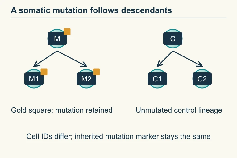 Original mutated and unmutated control lineage
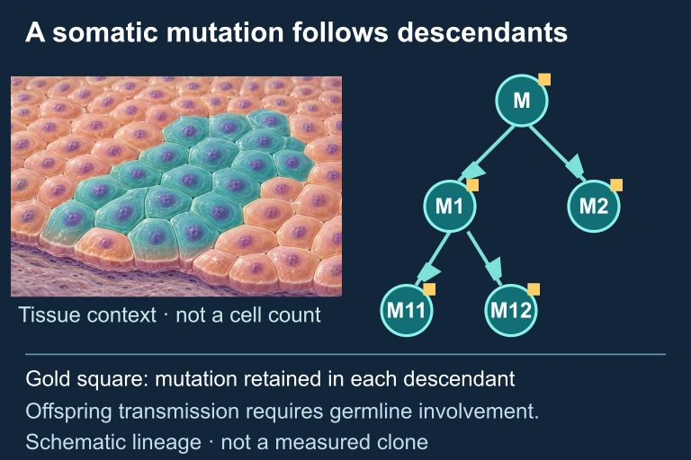 A somatic mutation can be inherited by descendant cells within a tissue without entering the next organismal generation. Illustration is schematic where scale or molecular structure is simplified.
SECTION 9
Practice Problems
Question 1: CONCEPTUAL
- A. Missense mutation, because the nucleotide sequence has changed
- B. Silent (synonymous) mutation, because the amino acid encoded remains the same
- C. Nonsense mutation, because the third codon position is the wobble position
- D. Frameshift mutation, because one nucleotide has been substituted
Show answer (B)
Question 2: BASIC
- A. A silent mutation because the protein still functions
- B. A nonsense mutation because it prevents drug binding
- C. A missense mutation that confers a gain-of-function (drug resistance)
- D. A frameshift mutation because the reading frame of rpoB is altered
Show answer (C)
Question 3: INTERMEDIATE
- A. The third position mutates at a higher rate than the first and second positions due to DNA polymerase infidelity at specific sites
- B. Substitutions at the third (wobble) position are more likely to be synonymous, and thus experience less purifying selection, allowing them to accumulate as neutral variation
- C. Positive selection favors substitutions at the third position because they produce novel amino acids beneficial for adaptation
- D. Mismatch repair enzymes preferentially correct errors at the first and second codon positions but not the third
Show answer (B)
Question 4: APPLIED
4 ptsShow answer
Question 5: CRITICAL THINKING
4 ptsShow answer
SUMMARY
Lesson Summary
Summary
A mutation is any permanent change in an organism's DNA nucleotide sequence. Mutations are classified by scale—point mutations (affecting one or a few nucleotides) versus chromosomal mutations (deletions, duplications, inversions, translocations, aneuploidy)—and by their effect on the protein product. Silent mutations exploit codon degeneracy and leave the amino acid sequence unchanged. Missense mutations substitute one amino acid for another, with consequences ranging from benign to lethal depending on the chemical properties of the substitution and its location in the protein. Nonsense mutations introduce premature stop codons, yielding truncated, typically nonfunctional proteins. Frameshift mutations (insertions or deletions not in multiples of three) corrupt the entire downstream reading frame and are usually the most severe.
Mutations arise through spontaneous errors during DNA replication (tautomeric shifts, depurination, deamination) and through induced mutagenesis by chemical, physical, or biological agents. Cells possess multi-layered defenses—proofreading and mismatch repair—that reduce the per-base error rate to approximately 10⁻⁹ to 10⁻¹⁰. Only germ-line mutations are heritable and contribute to evolutionary change; somatic mutations affect only the organism and its descendants of that cell lineage (e.g., cancer). Ultimately, mutation is the ultimate source of all genetic variation, providing the raw material upon which natural selection, genetic drift, and other evolutionary forces act to shape the diversity of life.