
SECTION 1
Historical Context & Motivation
The question of how substances enter and exit living cells has occupied biologists for well over a century. Early microscopists observed that cells could swell and shrink when exposed to different solutions, but the mechanism behind this selective permeability remained elusive. The gradual recognition that cells are bounded by a semipermeable membrane—a structure that permits some molecules to pass while excluding others—launched decades of research that converged on the modern understanding of membrane transport. This history reveals not just how scientific models evolve, but also why membrane transport is so fundamental: without it, cells could not regulate their internal environment, generate energy, or communicate with their surroundings.
1877 • Osmosis Described Quantitatively
1925 • Lipid Bilayer Proposed
1957 • Active Transport Characterized
1972 • Fluid Mosaic Model
2003 • Aquaporin Structure Resolved
These milestones converge on a central question that the AP Biology curriculum addresses directly: How do cells exploit the physical chemistry of membranes to control what enters and exits? Understanding the answer requires distinguishing between passive processes—driven by thermodynamic gradients—and active processes that require cellular energy input, a distinction that pervades every level of biological organization from individual enzyme kinetics to organ-level physiology.
SECTION 2
Core Principles of Membrane Transport
Membrane transport can be organized around a small set of foundational principles that distinguish the major categories of molecular movement. Every transport event depends on the physicochemical properties of the solute, the structure of the phospholipid bilayer, and the available energy sources. The plasma membrane's selective permeability arises because its hydrophobic interior excludes charged and large polar molecules, while small nonpolar molecules (O₂, CO₂) and water pass through with relative ease. Transport proteins embedded in the bilayer provide pathways for everything the lipid core rejects.
Passive Transport
Active Transport
Bulk Transport
Electrochemical Gradient
KEY TAKEAWAY
SECTION 3
Visual Overview of Membrane Transport
The diagram above captures the essential logic of membrane transport classification. On the far left, simple diffusion allows small, nonpolar molecules like O₂ and CO₂ to pass directly through the hydrophobic core of the bilayer without any protein assistance. Moving rightward, channel proteins provide hydrophilic tunnels for ions such as Na⁺, K⁺, and Cl⁻, while carrier proteins undergo conformational changes to shuttle larger polar molecules like glucose across the membrane. Both of these facilitated diffusion mechanisms are still passive—they follow the concentration gradient. The Na⁺/K⁺ pump (shown in pink) represents primary active transport, directly hydrolyzing ATP to move 3 Na⁺ out and 2 K⁺ in per cycle, generating an electrochemical gradient. Finally, endocytosis on the far right engulfs extracellular particles in membrane-derived vesicles—a form of bulk transport that also requires energy.
SECTION 4
Quantitative Framework & Mechanisms
While the AP Biology exam does not require complex mathematical derivations of transport equations, a quantitative understanding of the key relationships helps you predict the direction and rate of molecular movement. The fundamental driving force for passive transport is the concentration gradient, formalized in Fick's law of diffusion. For charged species, the Nernst equation and the concept of an electrochemical gradient become essential. Additionally, understanding osmolarity and water potential allows you to predict the direction of water flow in biological and experimental contexts.
FICK'S FIRST LAW (SIMPLIFIED)
Where J is the flux (amount per time), P is the permeability coefficient (related to diffusion coefficient and membrane thickness), A is the membrane surface area, and (C₂ − C₁) is the concentration difference across the membrane. The negative sign indicates net movement from high to low concentration.
WATER POTENTIAL
Water potential (Ψ) equals the sum of solute potential (Ψₛ) and pressure potential (Ψₚ). Solute potential is calculated as Ψₛ = −iCRT, where i = ionization constant, C = molar concentration, R = pressure constant (0.0831 L·bar/mol·K), and T = temperature in Kelvin. Water moves from high Ψ to low Ψ.
NA⁺/K⁺-ATPase STOICHIOMETRY
Each cycle of the sodium-potassium pump hydrolyzes one ATP to move 3 Na⁺ out and 2 K⁺ in. Because 3 positive charges exit and only 2 return, the pump is electrogenic—it contributes to the negative resting membrane potential.
AP Exam Tip
SECTION 5
Detailed Classification of Transport Types
A detailed breakdown of transport types is essential for the AP exam, which frequently tests whether students can identify specific mechanisms and predict outcomes under different conditions. The classification below distinguishes mechanisms by their energy requirements, the molecular players involved, and the biological contexts where each is most relevant.
| Feature | Simple Diffusion | Facilitated Diffusion | Active Transport |
|---|---|---|---|
| Energy source | None (ΔG < 0) | None (ΔG < 0) | ATP or ion gradient |
| Protein required? | No | Yes (channel or carrier) | Yes (pump) |
| Direction | Down gradient | Down gradient | Against gradient |
| Saturability | No (linear increase) | Yes (Vmax reached) | Yes (Vmax reached) |
| Specificity | Low (size & polarity) | High (substrate-specific) | High (substrate-specific) |
| Examples | O₂, CO₂, ethanol | Glucose (GLUT1), K⁺ leak channels | Na⁺/K⁺-ATPase, H⁺/sucrose symport |
Comparison of the three major categories of solute transport across biological membranes
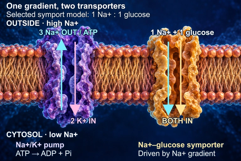 In this conceptual membrane, the Na+/K+ ATPase uses one ATP to move three Na+ out and two K+ in, maintaining a sodium electrochemical gradient. A separate symporter uses that gradient to bring Na+ and glucose inward; the 1:1 ratio is a selected teaching model and varies among transporters. The cutaways do not represent constitutively open pores: both transport cycles and the protein shapes are schematic. The juxtaposition does not assert a shared membrane face in a particular tissue.
SECTION 6
Worked Example: Water Potential Calculation
Water potential problems appear frequently on the AP Biology exam, particularly in free-response questions involving plant cells or dialysis tubing experiments. The following example walks through a complete calculation, demonstrating how to determine the direction of water movement between two compartments.
Predicting Direction of Osmosis in a Plant Cell
- 1
Step 1 — Identify Given Values
A plant cell with a solute concentration of 0.3 M sucrose (a nonionizing solute, so i = 1) is placed in a beaker of 0.1 M sucrose solution at 22 °C (295 K). The cell has a turgor pressure (Ψₚ) of 0.5 bar. The beaker is open, so its Ψₚ = 0. We need to determine the direction of net water movement.Ccell = 0.3 M, Cbeaker = 0.1 M, T = 295 K, Ψₚ(cell) = 0.5 bar, Ψₚ(beaker) = 0 - 2
Step 2 — Calculate Solute Potential (Ψₛ) for Each Compartment
Using the formula Ψₛ = −iCRT, where i = 1 (sucrose does not ionize), R = 0.0831 L·bar/(mol·K), and T = 295 K. For the cell: Ψₛ = −(1)(0.3)(0.0831)(295) = −7.35 bar. For the beaker: Ψₛ = −(1)(0.1)(0.0831)(295) = −2.45 bar.Ψₛ(cell) = −7.35 bar; Ψₛ(beaker) = −2.45 bar - 3
Step 3 — Calculate Total Water Potential (Ψ)
For the cell: Ψ = Ψₛ + Ψₚ = −7.35 + 0.5 = −6.85 bar. For the beaker: Ψ = Ψₛ + Ψₚ = −2.45 + 0 = −2.45 bar.Ψ(cell) = −6.85 bar; Ψ(beaker) = −2.45 bar - 4
Step 4 — Determine Direction of Water Movement
Water moves from regions of higher (less negative) water potential to regions of lower (more negative) water potential. The beaker has Ψ = −2.45 bar, which is higher than the cell's Ψ = −6.85 bar. Therefore, water will flow from the beaker into the cell. This makes biological sense: the cell has more solute (is hypertonic relative to the beaker), so water enters by osmosis. As water enters, turgor pressure increases until Ψ(cell) = Ψ(beaker), at which point equilibrium is reached and net water movement stops.Net water movement: beaker → cell (into the cell)
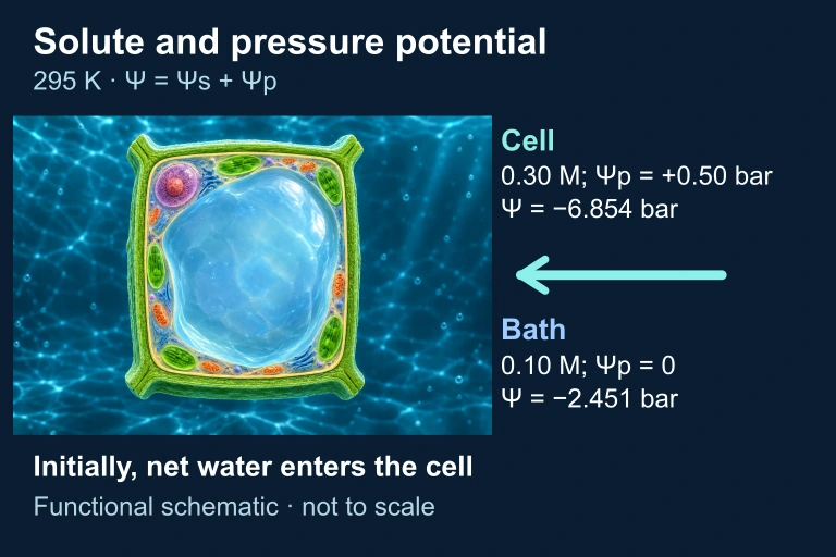 Turgor raises water potential and can oppose solute effects; these stated initial conditions still favor water entry. Values are an explicitly stated teaching model.
SECTION 7
Tonicity, Cell Responses & Common Confusions
One of the most testable areas on the AP exam involves predicting cellular responses in different osmotic environments. The concept of tonicity—the ability of an extracellular solution to cause water to move into or out of a cell—depends on the relative concentrations of nonpenetrating solutes on each side of the membrane. Critically, tonicity is distinct from osmolarity, because solutes that freely cross the membrane (like urea) contribute to osmolarity but not to tonicity, since they equilibrate and cannot sustain an osmotic gradient.
| Environment | Animal Cell Response | Plant Cell Response |
|---|---|---|
| Hypotonic (less solute outside) | Water enters → cell swells → may lyse (cytolysis) | Water enters → turgor pressure increases → cell becomes turgid (ideal state) |
| Isotonic (equal solute) | No net water movement → cell maintains normal shape | No net water movement → cell is flaccid (limp) |
| Hypertonic (more solute outside) | Water exits → cell shrinks (crenation) | Water exits → plasma membrane pulls from cell wall (plasmolysis) |
Cellular responses to different osmotic environments
COMMON EXAM PITFALL
Another frequently tested distinction involves primary versus secondary active transport. Primary active transport directly couples ATP hydrolysis to solute movement (e.g., the Na⁺/K⁺-ATPase). Secondary active transport (co-transport) uses the electrochemical gradient established by a primary pump to drive another solute against its gradient. For example, the Na⁺/glucose symporter in intestinal epithelial cells exploits the Na⁺ gradient created by the Na⁺/K⁺-ATPase to import glucose—a classic example of energy coupling across two transport events.
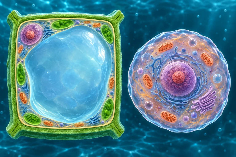 For nonpenetrating solutes, a plant wall limits expansion as water enters. Water loss retracts the protoplast from the wall during plasmolysis; animal cells lack this rigid wall.
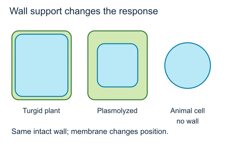 For nonpenetrating solutes, a plant wall limits expansion as water enters. Water loss retracts the protoplast from the wall during plasmolysis; animal cells lack this rigid wall.
SECTION 8
Connections to Signaling, Energetics & Disease
Membrane transport does not exist in isolation—it intersects with virtually every major topic in AP Biology. The proton motive force generated by the electron transport chain in mitochondria is itself an electrochemical gradient: protons (H⁺) are pumped across the inner mitochondrial membrane by complexes I, III, and IV, and their flow back through ATP synthase drives oxidative phosphorylation. Similarly, in chloroplasts, the thylakoid membrane maintains a steep H⁺ gradient that powers photosynthetic ATP synthesis. Understanding membrane transport therefore provides the mechanistic foundation for cellular energetics.
| AP Biology Topic | Connection to Membrane Transport |
|---|---|
| Cell Signaling | Ligand-gated ion channels open in response to neurotransmitter binding, enabling rapid signal transduction in neurons. The resting potential maintained by the Na⁺/K⁺-ATPase is prerequisite for action potentials. |
| Cellular Respiration | Chemiosmosis in the mitochondrial inner membrane: H⁺ ions pumped by ETC complexes flow down their gradient through ATP synthase, coupling transport to phosphorylation. |
| Photosynthesis | The thylakoid lumen accumulates H⁺ as water is split and electrons move through photosystems. This gradient drives ATP synthase in the chloroplast. |
| Immune System | Phagocytosis (a form of endocytosis) allows macrophages to engulf pathogens. Receptor-mediated endocytosis enables cells to internalize specific molecules like LDL cholesterol. |
| Cystic Fibrosis | Mutations in the CFTR gene disrupt a Cl⁻ channel protein, leading to thick mucus accumulation. This is a direct example of how a single transport protein defect causes systemic disease. |
How membrane transport connects to other AP Biology topics
Looking forward, upper-division courses in cell biology and biophysics treat membrane transport with mathematical rigor through the Goldman equation (which extends the Nernst equation to multiple ions), patch-clamp electrophysiology (which measures current through individual ion channels), and structural biology approaches that reveal the three-dimensional conformational changes of transporters at atomic resolution. The foundational principles covered here—gradient-driven movement, protein specificity, energy coupling, and electrochemical gradients—remain the conceptual backbone of all those advanced treatments.
SECTION 9
Practice Problems
Question 1: CONCEPTUAL
- A. Na⁺/K⁺-ATPase pumping sodium out of the cell
- B. Facilitated diffusion of glucose through GLUT1 transporters
- C. Exocytosis of neurotransmitter vesicles
- D. Phagocytosis of a bacterial cell
Show answer (B)
Question 2: BASIC CALCULATION
- A. −6.0 bar
- B. −4.0 bar
- C. −2.0 bar
- D. 0.0 bar
Show answer (C)
Question 3: INTERMEDIATE
- A. Simple diffusion with competitive inhibition
- B. Facilitated diffusion through a carrier protein that exhibits saturation and competitive inhibition
- C. Primary active transport that is inhibited by galactose binding to the pump
- D. Channel-mediated diffusion with allosteric regulation by galactose
Show answer (B)
Question 4: APPLIED
4 ptsShow answer
Question 5: CRITICAL THINKING
4 ptsShow answer
SUMMARY
Membrane Transport — Key Concepts Review
Summary
Membrane transport encompasses all mechanisms by which molecules cross the phospholipid bilayer. Passive transport (simple diffusion, facilitated diffusion, and osmosis) moves substances down their concentration or electrochemical gradient without energy input. Facilitated diffusion requires channel or carrier proteins and exhibits saturation kinetics and substrate specificity, distinguishing it from simple diffusion. Water potential (Ψ = Ψₛ + Ψₚ) predicts the direction of osmosis: water moves from higher to lower Ψ. Tonicity describes a solution's effect on cell volume and determines whether cells undergo lysis, crenation, turgidity, or plasmolysis.
Active transport moves substances against their gradient and requires energy. Primary active transport (e.g., Na⁺/K⁺-ATPase) directly hydrolyzes ATP, while secondary active transport (co-transport) harnesses an existing ion gradient. Bulk transport via endocytosis and exocytosis moves large particles in membrane-bound vesicles. These mechanisms connect to chemiosmosis in cellular respiration and photosynthesis, cell signaling via ion channels, and disease states such as cystic fibrosis caused by defective transport proteins.