AP Biology • Cells

Membrane Permeability

How the selective barrier of the plasma membrane governs molecular traffic to maintain cellular homeostasis.

AP Biology illustration with DNA, a microscope, and a leaf
Inside this lesson 10 sections 5 questions 8 figures

SECTION 1

Historical Context & Motivation

The concept of membrane permeability has roots stretching back to the earliest microscopic observations of cells and osmotic phenomena. Scientists knew that cells could maintain a distinct internal environment, yet the molecular basis of this selectivity remained elusive for decades. Understanding how lipid bilayers, proteins, and concentration gradients cooperate to regulate what enters and exits a cell became one of the central questions in cell biology. This pursuit ultimately revealed that membranes are not passive barriers but dynamic, selectively permeable structures that underpin virtually every physiological process, from nerve impulse transmission to nutrient absorption.

1831 • Osmosis Described

René Dutrochet formally described osmosis, demonstrating that water moves across semipermeable membranes in response to solute concentration differences — a phenomenon that implied cells possess a selective boundary.

1895 • Overton's Lipid Hypothesis

Charles Ernest Overton observed that nonpolar molecules cross cell membranes much more readily than polar or charged species, leading him to propose that the cell boundary is fundamentally lipid in nature.

1925 • Gorter & Grendel: Lipid Bilayer

Evert Gorter and François Grendel extracted lipids from red blood cells and showed that their surface area was roughly double the cell surface area, supporting the idea of a lipid bilayer structure.

1972 • Fluid Mosaic Model

S.J. Singer and Garth Nicolson proposed the fluid mosaic model, which described the membrane as a dynamic mosaic of phospholipids and proteins, providing the structural framework for understanding selective permeability.

2003 • Aquaporin Nobel Prize

Peter Agre received the Nobel Prize in Chemistry for discovering aquaporins — channel proteins that facilitate rapid water transport across membranes — demonstrating that even water requires protein-mediated pathways for high-flux movement.

Each of these milestones advanced a single overarching question: What determines which molecules can cross a biological membrane, and how does the cell regulate that traffic? The answer lies in the interplay between the hydrophobic core of the phospholipid bilayer, the specificity of transport proteins, and the thermodynamic gradients that drive molecular movement. Grasping membrane permeability is essential for understanding cellular homeostasis, signal transduction, and the pharmacological design of drugs that must cross biological membranes.

SECTION 2

Core Principles of Membrane Permeability

The plasma membrane's ability to discriminate among molecules rests on a few foundational principles. The phospholipid bilayer creates a hydrophobic interior that is intrinsically permeable to small nonpolar molecules but presents a formidable barrier to ions and large polar molecules. Embedded within this bilayer, transport proteins — channels and carriers — provide selective pathways that expand the membrane's functional permeability. The direction and rate of molecular movement are governed by concentration gradients, electrochemical gradients, and the input of metabolic energy (ATP). Together, these principles define selective permeability — the membrane's hallmark feature.

1

Hydrophobic Barrier

The nonpolar fatty acid tails of phospholipids form an interior that repels ions and polar molecules while allowing O₂, CO₂, and small hydrophobic molecules to diffuse freely.
2

Passive Transport

Movement down a concentration or electrochemical gradient without energy input. Includes simple diffusion, facilitated diffusion through channels, and osmosis.
3

Active Transport

Movement against a gradient, powered by ATP hydrolysis (primary) or by coupling to another ion's gradient (secondary/co-transport). Exemplified by the Na⁺/K⁺-ATPase.
4

Vesicular Transport

Endocytosis and exocytosis move macromolecules and bulk quantities of material across the membrane by packaging them in membrane-bound vesicles — bypassing the bilayer entirely.
5

Tonicity & Water Balance

The relative solute concentration outside versus inside the cell (hypertonic, hypotonic, isotonic) determines the net direction of water flow via osmosis, directly affecting cell volume and function.
✦

KEY TAKEAWAY

Think of the plasma membrane as a nightclub with a strict bouncer. Small, nonpolar molecules (VIPs) slip past the velvet rope without pausing. Polar molecules and ions must show a credential — a channel or carrier protein — to gain entry. Actively transported solutes are like guests who require the bouncer to physically escort them in against the crowd's flow, which costs the club energy (ATP). Macromolecules, too large for any door, are loaded into delivery vans (vesicles) that merge directly with the building wall. The bouncer's selectivity is what keeps the club's atmosphere precisely controlled — just as selective permeability maintains cellular homeostasis.

SECTION 3

Visualizing the Selectively Permeable Membrane

Selective Permeability of the Phospholipid BilayerExtracellular fluidCytoplasmHydrophobic core (fatty acid tails)ChannelCarrierO₂, CO₂(simple diffusion)Ions (Na⁺, K⁺)(facilitated – channel)Glucose(facilitated – carrier)Large polar / Ions ✗LegendPhospholipid head (hydrophilic)Hydrophobic tail regionChannel proteinCarrier proteinPermeable (passes through)Blocked (cannot cross unaided)
The phospholipid bilayer (purple heads, yellow interior) forms the selective barrier. Small nonpolar molecules like O₂ and CO₂ pass directly through by simple diffusion (green arrow). Ions such as Na⁺ and K⁺ require channel proteins (cyan), while glucose uses carrier proteins (pink). Large polar molecules and ions without a transporter are blocked (red X).

The diagram above illustrates the central theme of membrane permeability: the phospholipid bilayer is not uniformly impermeable — its selectivity depends on the physical and chemical properties of each solute. Three properties determine a molecule's ability to cross: size, polarity, and charge. Small, uncharged, nonpolar molecules — such as O₂, CO₂, and N₂ — dissolve into the hydrophobic core and pass through readily. Water, despite being polar, is small enough to cross slowly by simple diffusion, though cells dramatically increase water flux via aquaporin channels. Larger polar molecules like glucose and amino acids require specific carrier proteins, and ions are almost completely excluded from the bilayer unless they pass through ion channels or are actively pumped.

SECTION 4

Transport Mechanisms in Depth

While much of membrane permeability is qualitative in AP Biology, a quantitative understanding of diffusion and osmosis deepens conceptual insight. Fick's law of diffusion provides the mathematical framework linking the rate of diffusion to measurable variables, and the concept of water potential (Ψ) formalizes the prediction of osmotic water movement — a concept tested directly on the AP exam.

FICK'S LAW OF DIFFUSION

J=−P×A×(C2−C1)J = −P × A × (C₂ − C₁)

J = net flux (mol·s⁻¹); P = permeability coefficient (cm·s⁻¹), which depends on the molecule's partition coefficient and the membrane thickness; A = membrane surface area; C₂ − C₁ = concentration difference across the membrane. The negative sign indicates net movement from high to low concentration.

WATER POTENTIAL (AP BIOLOGY)

Ψ=Ψs+ΨpΨ = Ψₛ + Ψₚ

Ψ = water potential (bars or MPa); Ψₛ = solute potential (always ≤ 0 for a solution); Ψₚ = pressure potential (positive in turgid plant cells, zero in open containers). Water moves from regions of higher Ψ to regions of lower Ψ.

SOLUTE POTENTIAL

Ψs=−iCRTΨₛ = −iCRT

i = ionization constant (van 't Hoff factor); C = molar concentration of solute; R = pressure constant (0.0831 L·bar·mol⁻¹·K⁻¹); T = temperature in Kelvin. More solute → more negative Ψₛ → lower water potential.

These equations reinforce a key conceptual point: the direction of molecular movement is always dictated by gradients. For non-water solutes, the concentration gradient (and for ions, the additional electrical gradient forming the electrochemical gradient) determines the direction and rate of passive transport. For water, Ψ is the master variable. Active transport, by contrast, uses metabolic energy to move solutes against their gradient — analogous to pumping water uphill.

💡

AP EXAM TIP

Free-response questions frequently ask you to predict the direction of water movement given solute concentrations on either side of a membrane. Always calculate Ψ for each compartment, then state that water moves from the side with higher Ψ to the side with lower Ψ. Remember: adding solute makes Ψₛ more negative, which lowers the overall Ψ.

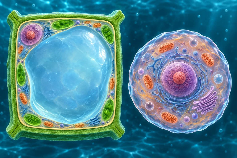
Plant wall is thick and distinct from inner membrane; large central vacuole and peripheral cytoplasm visible. Animal cell has flexible boundary and no wall. Separate exact panel: Paired plant states retain the same wall while the inner membrane is pressed outward or retracted; animal membrane has no surrounding wall.For nonpenetrating solutes, a plant wall limits expansion as water enters. Water loss retracts the protoplast from the wall during plasmolysis; animal cells lack this rigid wall.
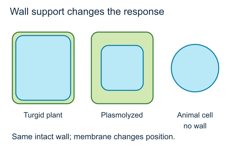
Wall support changes the response. For nonpenetrating solutes, a plant wall limits expansion as water enters. Water loss retracts the protoplast from the wall during plasmolysis; animal cells lack this rigid wall.For nonpenetrating solutes, a plant wall limits expansion as water enters. Water loss retracts the protoplast from the wall during plasmolysis; animal cells lack this rigid wall.

SECTION 5

Classifying Transport Across Membranes

Classification of Membrane TransportMembrane TransportPassive (no ATP)Active (requires ATP)Simple DiffusionO₂, CO₂, steroidsNo protein neededFacilitated DiffusionGlucose (GLUT), ionsChannel or carrierOsmosisWater via bilayeror aquaporinsPrimary ActiveNa⁺/K⁺-ATPaseDirect ATP hydrolysisSecondary ActiveSGLT (glucose-Na⁺)Coupled to ion gradientVesicular (Bulk)Endocytosis / ExocytosisMacromolecules, particlesKey DistinctionPassive transport: moves DOWN the gradient (ΔG < 0, spontaneous). Active transport: moves AGAINST the gradient (ΔG > 0, requires energy input).
This flowchart categorizes membrane transport into passive (green border, no ATP) and active (red border, requires ATP). Under passive transport, simple diffusion, facilitated diffusion, and osmosis are distinguished by their need for protein assistance. Active transport includes primary (direct ATP use), secondary (coupled ion gradients), and vesicular (endocytosis/exocytosis) mechanisms.
FeatureSimple DiffusionFacilitated DiffusionActive Transport
DirectionDown gradientDown gradientAgainst gradient
Protein RequiredNoYes (channel or carrier)Yes (pump or coupled transporter)
Energy (ATP)Not requiredNot requiredRequired
Saturation KineticsNo (linear rate)Yes (V_max plateau)Yes (V_max plateau)
SpecificityLow (depends on lipid solubility)High (protein-specific)High (protein-specific)
ExamplesO₂, CO₂, ethanol, steroid hormonesGlucose (GLUT1), K⁺ leak channels, aquaporinsNa⁺/K⁺-ATPase, H⁺ pump, SGLT1 (secondary)

Comparison of the three major transport mechanisms across biological membranes

A critical distinction highlighted in the table concerns saturation kinetics. Simple diffusion increases linearly with the concentration gradient because it does not depend on a finite number of protein binding sites. Facilitated diffusion and active transport, however, exhibit a maximum rate (Vmax) because all available transport proteins become occupied at high substrate concentrations. This concept parallels enzyme kinetics and is a favorite source of AP exam graph-interpretation questions: if a transport rate versus concentration curve plateaus, the process is protein-mediated.
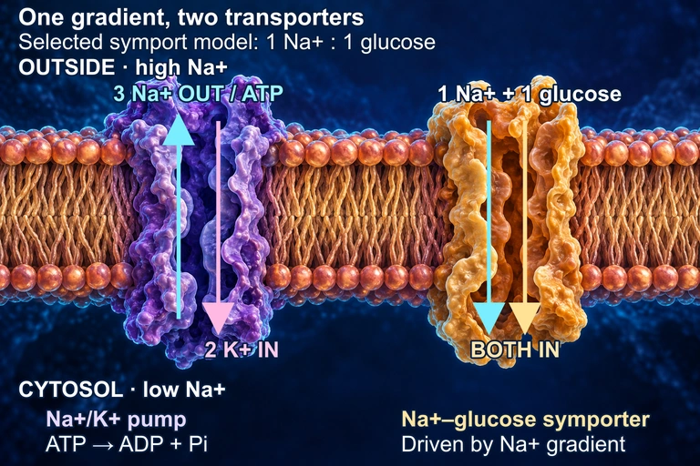
A conceptual membrane has higher sodium outside and lower sodium in the cytosol. A violet pump moves three sodium out and two potassium in per ATP. A separate gold symporter brings one sodium and one glucose inward, using the gradient. Protein forms and their juxtaposition are schematic.In this conceptual membrane, the Na+/K+ ATPase uses one ATP to move three Na+ out and two K+ in, maintaining a sodium electrochemical gradient. A separate symporter uses that gradient to bring Na+ and glucose inward; the 1:1 ratio is a selected teaching model and varies among transporters. The cutaways do not represent constitutively open pores: both transport cycles and the protein shapes are schematic. The juxtaposition does not assert a shared membrane face in a particular tissue.

SECTION 6

Worked Example: Predicting Osmotic Water Movement

A common AP Biology free-response task asks you to calculate water potential and predict the direction of osmosis. The following example walks through this process step by step, using the water potential equation Ψ = Ψₛ + Ψₚ.

Predicting Water Flow Between Two Compartments

  1. 1

    Step 1 — Identify Given Values

    An open beaker (Compartment A) contains a 0.4 M sucrose solution at 27 °C. A plant cell (Compartment B) has an internal sucrose concentration of 0.6 M and a turgor pressure of 3.0 bars. The ionization constant for sucrose (a non-electrolyte) is i = 1.
    CAC_A = 0.4 M, CBC_B = 0.6 M, Ψₚ(A) = 0 bars, Ψₚ(B) = 3.0 bars, T = 300 K, R = 0.0831 L·bar·mol⁻¹·K⁻¹
  2. 2

    Step 2 — Calculate Ψₛ for Each Compartment

    Apply Ψₛ = −iCRT for each compartment. For Compartment A: Ψₛ = −(1)(0.4)(0.0831)(300) = −9.97 bars ≈ −9.97 bars. For Compartment B: Ψₛ = −(1)(0.6)(0.0831)(300) = −14.96 bars ≈ −14.96 bars.
    Ψₛ(A) = −9.97 bars; Ψₛ(B) = −14.96 bars
  3. 3

    Step 3 — Calculate Total Ψ for Each Compartment

    Total water potential Ψ = Ψₛ + Ψₚ. For Compartment A (open beaker): Ψ(A) = −9.97 + 0 = −9.97 bars. For Compartment B (plant cell): Ψ(B) = −14.96 + 3.0 = −11.96 bars.
    Ψ(A) = −9.97 bars; Ψ(B) = −11.96 bars
  4. 4

    Step 4 — Predict the Direction of Water Movement

    Water moves from regions of higher Ψ to regions of lower Ψ. Since Ψ(A) = −9.97 bars is greater (less negative) than Ψ(B) = −11.96 bars, net water movement occurs from Compartment A (beaker) into Compartment B (the plant cell). As water enters the cell, turgor pressure will increase until equilibrium is reached.
    Water moves from the beaker (Ψ = −9.97 bars) into the plant cell (Ψ = −11.96 bars).

SECTION 7

Factors Affecting Permeability & Limitations of Simple Models

The permeability of a biological membrane is not fixed — it varies with composition, temperature, and cellular context. Several factors modulate how readily molecules cross the bilayer, and understanding these factors is essential for interpreting experimental data on the AP exam.

FactorEffect on PermeabilityMechanism / Explanation
Temperature ↑Increases permeabilityGreater kinetic energy increases membrane fluidity and molecular diffusion rates.
CholesterolBuffers fluidityAt high temperatures, cholesterol restrains phospholipid movement (decreases permeability). At low temperatures, it prevents tight packing (increases permeability). Acts as a fluidity buffer.
Unsaturated fatty acid tailsIncreases permeabilityKinks from cis double bonds prevent tight packing, creating more space in the hydrophobic core.
Saturated fatty acid tailsDecreases permeabilityStraight tails pack closely, creating a more rigid and less permeable bilayer.
Number of transport proteinsIncreases facilitated & active transport ratesMore channels or pumps raise V_max, allowing greater solute flux at saturation.
Molecular size & polaritySmaller / nonpolar = more permeableSmall, nonpolar molecules dissolve readily into the hydrophobic core; large, charged species cannot.

Factors modulating biological membrane permeability
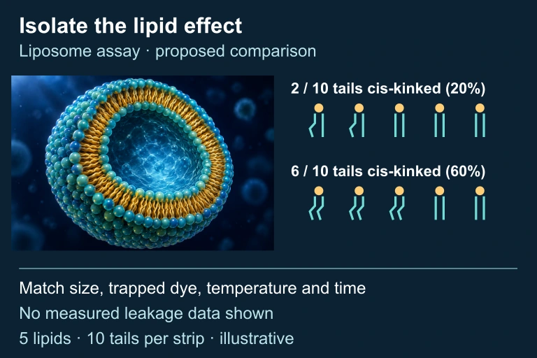
A contextual liposome accompanies two strips of five two-tailed phospholipids. The low strip has two cis-kinked tails out of ten; the high strip has six out of ten. The companion has equal vesicles with ten gold dye markers each and the same exact tail counts.Illustrative lipid-composition controls compare 2 / 10 tails cis-kinked (20%) with 6 / 10 tails cis-kinked (60%). Every strip has five two-tailed lipids. Vesicle size, trapped dye, temperature and time are matched; no measured leakage data are shown.
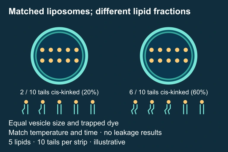
A contextual liposome accompanies two strips of five two-tailed phospholipids. The low strip has two cis-kinked tails out of ten; the high strip has six out of ten. The companion has equal vesicles with ten gold dye markers each and the same exact tail counts.Illustrative lipid-composition controls compare 2 / 10 tails cis-kinked (20%) with 6 / 10 tails cis-kinked (60%). Every strip has five two-tailed lipids. Vesicle size, trapped dye, temperature and time are matched; no measured leakage data are shown.

✦

KEY TAKEAWAY

Simple permeability models treat the membrane as a uniform hydrophobic slab, but real membranes are heterogeneous mosaics. Lipid rafts (cholesterol-enriched microdomains), the glycocalyx, and cytoskeletal attachments all influence local permeability. For the AP exam, the critical simplification is valid — but for advanced studies, recognize that membrane heterogeneity is the norm, not the exception. Think of it like weather forecasting: a simple model (temperature predicts snow versus rain) works most of the time, but micro-climates (valleys, coastlines) create local exceptions that require more refined models.

SECTION 8

Connections to Signal Transduction & Pharmacology

Membrane permeability is not an isolated concept — it integrates deeply with signal transduction, neurobiology, and drug design. The AP Biology curriculum connects membrane transport to several Big Ideas, including the role of ligand-gated ion channels in synaptic signaling and the importance of membrane receptor proteins in cell communication. Understanding how permeability is dynamically regulated — through gated channels that open or close in response to voltage changes, ligand binding, or mechanical stimuli — is the gateway to neuroscience and endocrinology.

ConceptAP Biology LevelAdvanced / College Extension
Ion channelsFacilitate passive ion movement; can be gatedPatch-clamp electrophysiology; single-channel conductance measurements; channelopathies (e.g., cystic fibrosis)
Membrane potentialResting potential maintained by Na⁺/K⁺-ATPase and K⁺ leak channelsNernst equation and Goldman equation quantify equilibrium and resting potentials; action potential dynamics
Drug deliveryLipid-soluble drugs cross membranes more easilyLipinski's Rule of Five predicts oral bioavailability; liposome-based drug carriers; nanoparticle delivery across the blood-brain barrier
Water potentialΨ = Ψₛ + Ψₚ predicts direction of osmosisSoil-plant-atmosphere continuum modeled with water potential; clinical osmolarity and IV fluid design

AP-level versus advanced extensions of membrane permeability concepts

As you move into college biology, biochemistry, or physiology, the qualitative permeability rules you learn in AP Biology become quantitative tools — the Nernst equation for ion equilibrium potentials, the Goldman equation for membrane potential, and the Michaelis-Menten framework for transporter kinetics. Mastering the conceptual foundations now will make those advanced treatments far more intuitive.

SECTION 9

Practice Problems

Question 1: CONCEPTUAL

A researcher places red blood cells in three different solutions and observes that in Solution X the cells swell and lyse, in Solution Y they remain unchanged, and in Solution Z they shrink (crenate). Which of the following correctly identifies the tonicity of each solution relative to the cytoplasm of the red blood cells?
  • A. X = hypertonic; Y = isotonic; Z = hypotonic
  • B. X = hypotonic; Y = isotonic; Z = hypertonic
  • C. X = isotonic; Y = hypertonic; Z = hypotonic
  • D. X = hypotonic; Y = hypertonic; Z = isotonic
Show answer (B)
The correct answer is B. In a hypotonic solution (X), the extracellular solute concentration is lower than the cytoplasm, so water enters the cell by osmosis, causing swelling and lysis. In an isotonic solution (Y), solute concentrations are equal on both sides, producing no net water movement. In a hypertonic solution (Z), the extracellular solute concentration exceeds the intracellular concentration, so water leaves the cell, causing it to shrink (crenate). Choice A reverses X and Z. Choice C incorrectly assigns X as isotonic when lysis clearly indicates net water gain. Choice D incorrectly identifies Y and Z.

Question 2: BASIC CALCULATION

A plant cell is placed in an open beaker containing a 0.3 M NaCl solution at 22 °C (295 K). NaCl fully dissociates (i = 2). What is the solute potential (Ψₛ) of the solution? (R = 0.0831 L·bar·mol⁻¹·K⁻¹)
  • A. −7.35 bars
  • B. −14.70 bars
  • C. −4.91 bars
  • D. −24.50 bars
Show answer (B)
The correct answer is B. Using Ψₛ = −iCRT: Ψₛ = −(2)(0.3)(0.0831)(295) = −(2)(0.3)(24.51) = −14.71 bars, which rounds to −14.70 bars. Choice A (−7.35 bars) results from using i = 1 instead of 2, forgetting that NaCl dissociates into Na⁺ and Cl⁻. Choice C (−4.91 bars) results from using i = 1 and dividing by an additional factor, an arithmetic error. Choice D (−24.50 bars) likely results from using an incorrect temperature or doubling the concentration instead of using the ionization constant.

Question 3: INTERMEDIATE

A student measures the rate of glucose uptake by cells at increasing external glucose concentrations and generates a curve that plateaus at high concentrations. When the student adds a metabolic poison that depletes ATP, the glucose uptake rate remains unchanged. Which type of transport best explains the observed glucose uptake?
  • A. Simple diffusion through the phospholipid bilayer
  • B. Primary active transport via a glucose-specific ATPase
  • C. Facilitated diffusion via a glucose carrier protein
  • D. Secondary active transport coupled to a Na⁺ gradient
Show answer (C)
The correct answer is C. Two observations must be explained: (1) the rate plateaus, indicating a protein-mediated process with saturation kinetics, and (2) the process is ATP-independent, since the metabolic poison did not affect uptake. Facilitated diffusion through a glucose carrier (e.g., GLUT1) satisfies both criteria — it uses a finite number of carriers (plateau at V_max) and requires no ATP. Choice A is wrong because simple diffusion through the bilayer would show a linear, non-saturating relationship and glucose is too large and polar to cross efficiently without a protein. Choice B is wrong because primary active transport requires direct ATP hydrolysis, which was inhibited. Choice D is wrong because secondary active transport depends on an ion gradient maintained by an ATP-dependent pump (Na⁺/K⁺-ATPase), so depleting ATP would eventually collapse the gradient and reduce uptake.

Question 4: APPLIED

4 pts
A student hypothesizes that increasing the proportion of unsaturated fatty acids in the cell membrane increases the rate of simple diffusion of ethanol across the membrane. Design a controlled experiment to test this hypothesis using artificial phospholipid vesicles (liposomes). In your response: (a) Identify the independent variable, dependent variable, and at least two controlled (standardized) variables. (b) Describe the experimental setup, including a negative control. (c) Predict the expected results if the hypothesis is supported. (d) Explain the biological reasoning behind your prediction.
Show answer
Rubric (4 points): (a) Variables (1 point): The independent variable is the proportion of unsaturated fatty acids in the liposome bilayer (e.g., 0%, 25%, 50%, 75%, 100% unsaturated phospholipids). The dependent variable is the rate of ethanol diffusion across the liposome membrane, measured as the concentration of ethanol appearing inside the vesicles over time. Controlled variables include total phospholipid concentration, liposome diameter, ethanol concentration in the external medium, temperature, and pH. (b) Experimental setup (1 point): Prepare five sets of liposomes with differing ratios of saturated to unsaturated phospholipids (e.g., 100:0, 75:25, 50:50, 25:75, 0:100). Suspend each set in an identical ethanol solution. At timed intervals, sample and measure ethanol concentration inside the vesicles (e.g., via gas chromatography or spectrophotometry). The 100% saturated liposome group (0% unsaturated) serves as a negative control — the baseline for minimal bilayer fluidity. (c) Predicted results (1 point): If the hypothesis is supported, liposomes with higher proportions of unsaturated fatty acids will show faster ethanol accumulation over time, producing a steeper uptake curve. The 100% unsaturated group should reach equilibrium fastest, and the 100% saturated group should reach equilibrium slowest. (d) Biological reasoning (1 point): Unsaturated fatty acid tails contain cis double bonds that introduce kinks, preventing tight packing of the phospholipid tails. This increases membrane fluidity and creates transient gaps in the hydrophobic core, allowing small molecules like ethanol to partition into and diffuse across the bilayer more rapidly.

Question 5: CRITICAL THINKING

4 pts
The following data were collected for four molecules tested for their ability to cross an artificial phospholipid bilayer (no proteins present) at 25 °C: Molecule A: MW = 32, charge = 0, log partition coefficient = 0.7 → relative permeability = 100 Molecule B: MW = 180, charge = 0, log partition coefficient = −3.2 → relative permeability = 0.01 Molecule C: MW = 44, charge = 0, log partition coefficient = 1.2 → relative permeability = 135 Molecule D: MW = 23, charge = +1, log partition coefficient = −3.8 → relative permeability = 0.001 (a) Identify which physical/chemical property is most important in determining a molecule's permeability through a pure lipid bilayer. Use evidence from the data to support your claim. (b) Explain why Molecule D has the lowest permeability despite having the smallest molecular weight. (c) Predict what would happen to the relative permeability of Molecule B if the bilayer were embedded with GLUT1 glucose transporters. Justify your prediction. (d) A pharmaceutical company wants to design a drug that crosses cell membranes easily without a carrier protein. Based on the data, recommend two molecular characteristics the drug should have, and explain your reasoning.
Show answer
Rubric (4 points): (a) Key property identification (1 point): The log partition coefficient (a measure of hydrophobicity/lipid solubility) is the most important determinant of bilayer permeability. Evidence: Molecules A and C have positive log partition coefficients and high permeability (100 and 135, respectively), while Molecules B and D have strongly negative log partition coefficients and extremely low permeability (0.01 and 0.001). The correlation between higher partition coefficients and higher permeability, across a range of molecular weights, demonstrates that lipid solubility is the dominant factor. (b) Molecule D explanation (1 point): Despite being the smallest molecule (MW = 23), Molecule D carries a +1 charge. Charged species are repelled by the hydrophobic interior of the phospholipid bilayer because the energy cost of moving an ion from an aqueous environment into a nonpolar medium is extremely high (the Born energy barrier). This charge effect overwhelms any advantage conferred by small size, as reflected in its very negative log partition coefficient (−3.8). (c) Prediction for Molecule B with GLUT1 (1 point): Molecule B (MW = 180, uncharged, log partition coefficient = −3.2) matches the profile of glucose. If GLUT1 transporters are embedded in the bilayer, the relative permeability of Molecule B would increase dramatically because GLUT1 provides a hydrophilic pathway through the membrane, bypassing the hydrophobic core. The transport rate would now depend on GLUT1 density and would show saturation kinetics rather than the linear rate expected for simple diffusion. (d) Drug design recommendation (1 point): The drug should (1) be small in molecular weight (ideally < 500 Da, consistent with Lipinski's Rule of Five) to transit the bilayer efficiently, and (2) have a positive log partition coefficient (moderate hydrophobicity, nonpolar character) so it can dissolve into and cross the hydrophobic core. It should be uncharged at physiological pH, since charged species are effectively excluded from the lipid interior, as demonstrated by Molecule D's extremely low permeability.

SUMMARY

Membrane Permeability — Key Concepts Review

Summary

The phospholipid bilayer establishes a selectively permeable barrier whose permeability depends on a molecule's size, polarity, and charge. Small nonpolar molecules cross by simple diffusion, while polar molecules and ions require transport proteins — channels for facilitated diffusion (passive, down the gradient) or pumps for active transport (ATP-dependent, against the gradient). Macromolecules use vesicular transport (endocytosis/exocytosis).

For the AP exam, master the water potential equation (Ψ = Ψₛ + Ψₚ) and the solute potential formula (Ψₛ = −iCRT) to predict the direction of osmotic water movement. Remember that membrane fluidity — modulated by cholesterol, fatty acid saturation, and temperature — directly affects how readily solutes permeate. Protein-mediated transport shows saturation kinetics (a plateau on a rate vs. concentration graph), distinguishing it from simple diffusion on AP exam data-interpretation questions.

Varsity Tutors • AP Biology • Membrane Permeability