
SECTION 1
Historical Context & Motivation
The story of membrane transport begins with the realization that cells are not merely passive containers but dynamic systems that carefully regulate the passage of molecules across their boundaries. Early microscopists observed that cells could swell and shrink in solutions of varying concentrations, but the molecular explanation for these phenomena remained elusive for centuries. The development of the fluid mosaic model of the cell membrane in the twentieth century provided the structural framework necessary to understand how transport occurs. Today, our understanding of membrane transport integrates principles from thermodynamics, protein biochemistry, and cell biology, revealing that the selective permeability of biological membranes is one of the most fundamental requirements for life.
1748 • Osmosis Described
1877 • Osmotic Pressure Quantified
1925 • Lipid Bilayer Proposed
1972 • Fluid Mosaic Model
2003 • Aquaporin Nobel Prize
These historical milestones converge on a central biological question: How do cells control which substances enter and exit, and what energy sources drive these movements? Answering this question requires understanding the interplay between the thermodynamics of diffusion, the architecture of the phospholipid bilayer, and the specificity of membrane transport proteins. These mechanisms are not merely academic — they underpin nerve impulses, nutrient absorption, kidney filtration, and virtually every physiological process in living organisms.
SECTION 2
Core Principles of Membrane Transport
Membrane transport can be broadly classified by two criteria: whether the process requires metabolic energy and whether it involves membrane-bound proteins. All transport mechanisms are governed by the second law of thermodynamics — substances spontaneously move down their concentration (or electrochemical) gradients unless energy is expended to move them against those gradients. The phospholipid bilayer acts as a selectively permeable barrier: small, nonpolar molecules like O2 and CO2 pass through readily, whereas ions, large polar molecules, and macromolecules generally cannot cross without assistance.
Passive Transport
Active Transport
Bulk Transport
Selective Permeability
KEY TAKEAWAY
SECTION 3
Visual Overview of Transport Mechanisms
The diagram above emphasizes a critical organizational principle: the distinction between passive and active mechanisms is rooted in thermodynamics, specifically whether the free energy change (ΔG) of the transport process is negative (spontaneous) or positive (requires energy input). In passive transport, molecules move from regions of higher concentration to regions of lower concentration, increasing entropy and releasing free energy. In active transport, cells must couple an energetically unfavorable process — moving molecules against their gradient — with an exergonic reaction such as ATP hydrolysis. The proteins embedded in the bilayer (channels, carriers, and pumps) provide the structural specificity that allows cells to control which substances cross and at what rate, a hallmark of selective permeability.
SECTION 4
Thermodynamic & Quantitative Framework
Understanding membrane transport at a deeper level requires connecting the biological observations to thermodynamic principles. The free energy of transporting an uncharged solute across a membrane depends on the ratio of concentrations on either side. For charged ions, the electrical potential difference across the membrane (membrane potential) also contributes, giving rise to the electrochemical gradient. Two key quantitative relationships govern these processes.
FICK'S LAW OF DIFFUSION (SIMPLIFIED)
Where J = net flux (mol/s), P = permeability coefficient (cm/s), A = membrane surface area, C₁ and C₂ = concentrations on each side of the membrane. Net flux is proportional to the concentration difference and the membrane area, and the negative sign indicates movement from high to low concentration.
FREE ENERGY OF TRANSPORT (UNCHARGED SOLUTE)
Where R = gas constant (8.314 J/mol·K), T = absolute temperature in Kelvin, and C_in / C_out = the ratio of solute concentration inside vs. outside the cell. When ΔG < 0, transport is thermodynamically favorable (passive); when ΔG > 0, energy input is required (active).
WATER POTENTIAL (BIOLOGY-SPECIFIC)
Where Ψ = water potential (in bars or MPa), Ψ_s = solute potential (always ≤ 0; more solute = more negative), and Ψ_p = pressure potential (positive in turgid plant cells, zero in animal cells under standard conditions). Water moves from regions of higher Ψ to regions of lower Ψ.
AP Exam Tip
SECTION 5
Detailed Classification of Transport Types
A thorough classification of transport mechanisms requires distinguishing among their energy requirements, protein involvement, direction relative to gradients, and the types of substances they move. The following table and diagram organize these distinctions systematically, providing the level of detail expected on the AP Biology exam.
| Mechanism | Energy Source | Protein Required? | Direction | Example Molecules |
|---|---|---|---|---|
| Simple Diffusion | None (ΔG < 0) | No | Down gradient | O₂, CO₂, ethanol, steroid hormones |
| Osmosis | None (ΔG < 0) | Aquaporins (optional) | High Ψ → Low Ψ | H₂O |
| Facilitated Diffusion (channels) | None (ΔG < 0) | Yes — channel proteins | Down gradient | Na⁺, K⁺, Cl⁻ through gated ion channels |
| Facilitated Diffusion (carriers) | None (ΔG < 0) | Yes — carrier proteins | Down gradient | Glucose (GLUT transporters), amino acids |
| Primary Active Transport | ATP hydrolysis | Yes — ATPase pumps | Against gradient | Na⁺/K⁺-ATPase, Ca²⁺-ATPase, H⁺ pump |
| Secondary Active Transport | Ion gradient (indirect ATP) | Yes — cotransporters | Against gradient (coupled) | Na⁺/glucose symporter, Na⁺/H⁺ antiporter |
| Endocytosis | ATP | Yes — receptor/cytoskeletal | Into cell | Bacteria (phagocytosis), LDL (receptor-mediated) |
| Exocytosis | ATP | Yes — SNARE proteins | Out of cell | Neurotransmitters, hormones, mucus |
Comprehensive classification of membrane transport mechanisms
The distinction between channel proteins and carrier proteins is frequently tested on the AP Biology exam. Channel proteins form aqueous pores through which specific ions or small molecules can flow at very high rates (up to 10⁸ ions per second) and can be gated — opened or closed in response to voltage, ligands, or mechanical stress. Carrier proteins, by contrast, bind their substrate on one side of the membrane, undergo a conformational change, and release it on the other side; this process is slower (10²–10⁴ molecules per second) but allows for greater specificity. Both channels and carriers can mediate passive facilitated diffusion, but only carriers (not channels) participate in active transport, because the conformational change can be coupled to energy input.
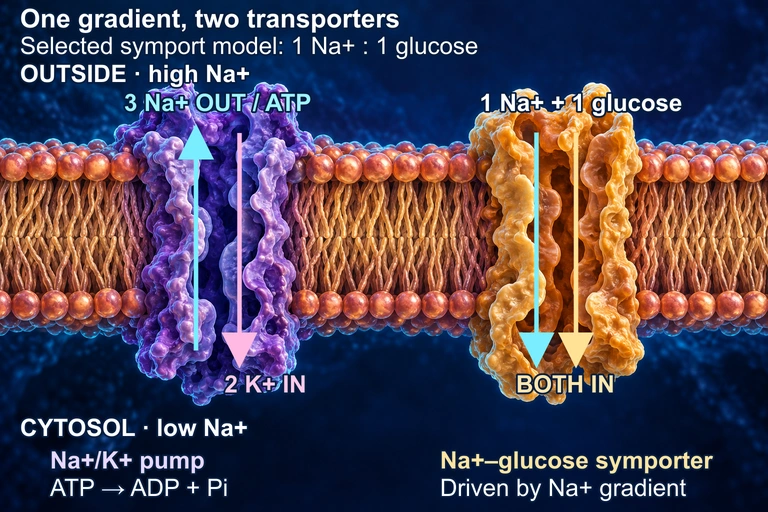 In this conceptual membrane, the Na+/K+ ATPase uses one ATP to move three Na+ out and two K+ in, maintaining a sodium electrochemical gradient. A separate symporter uses that gradient to bring Na+ and glucose inward; the 1:1 ratio is a selected teaching model and varies among transporters. The cutaways do not represent constitutively open pores: both transport cycles and the protein shapes are schematic. The juxtaposition does not assert a shared membrane face in a particular tissue.
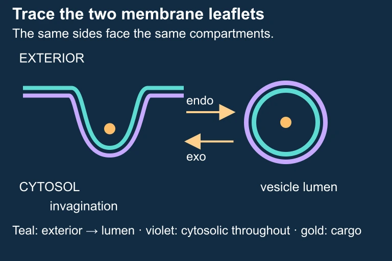 Trace the two membrane leaflets through vesicle formation and fusion.
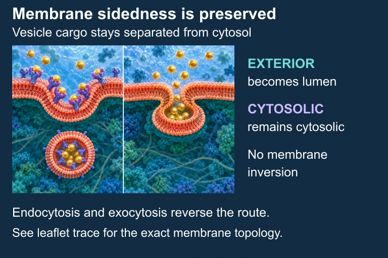 Vesicle budding and fusion preserve which membrane surface faces cytosol and which faces the lumen or exterior. Illustration is schematic where scale or molecular structure is simplified.
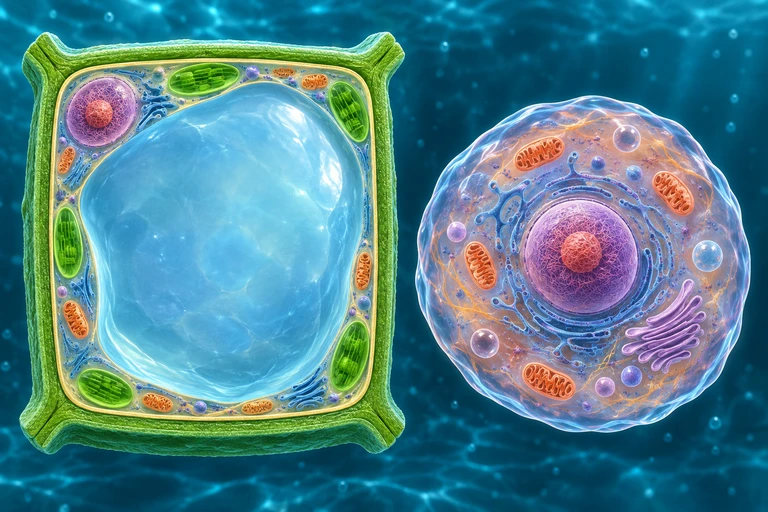 For nonpenetrating solutes, a plant wall limits expansion as water enters. Water loss retracts the protoplast from the wall during plasmolysis; animal cells lack this rigid wall.
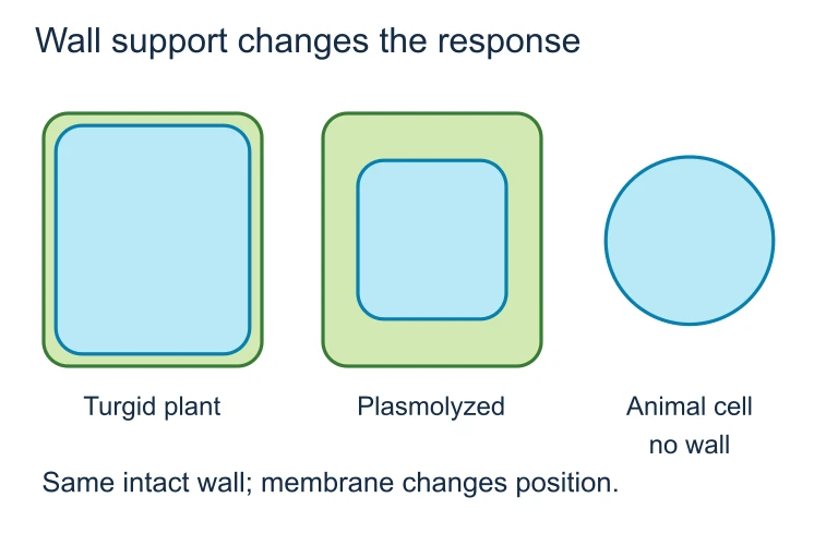 For nonpenetrating solutes, a plant wall limits expansion as water enters. Water loss retracts the protoplast from the wall during plasmolysis; animal cells lack this rigid wall.
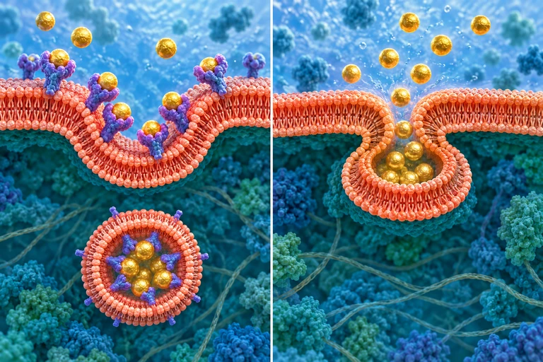 Endocytosis encloses extracellular cargo in a vesicle lumen; exocytosis opens that lumen to the extracellular space. Membrane fusion and budding preserve which membrane side faces the cargo.
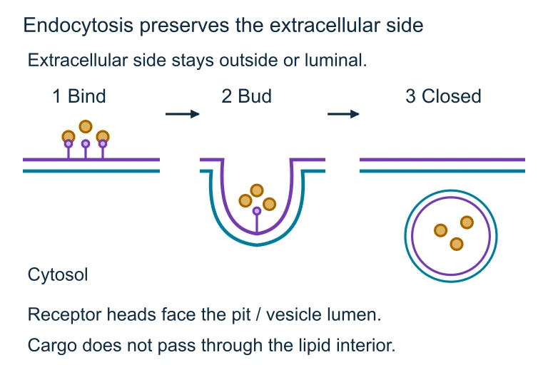 Endocytosis encloses extracellular cargo in a vesicle lumen; exocytosis opens that lumen to the extracellular space. Membrane fusion and budding preserve which membrane side faces the cargo.
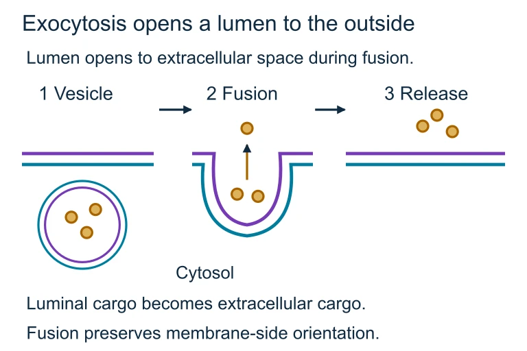 During exocytosis, the vesicle lumen opens to the extracellular space while membrane-side orientation is preserved.
SECTION 6
Worked Example: Water Potential & Osmosis
Water potential calculations are a staple of the AP Biology exam. The following example demonstrates how to determine the direction and extent of water movement between a plant cell and its surrounding solution.
Predicting Water Movement in a Plant Cell
- 1
Step 1 — Identify Given Values
A plant cell with a solute potential (Ψs) of −0.9 MPa and a pressure potential (Ψp) of +0.4 MPa is placed in a beaker of sucrose solution with Ψs = −0.3 MPa and Ψp = 0 MPa (open container, no pressure).Cell: Ψs = −0.9 MPa, Ψp = +0.4 MPa; Solution: Ψs = −0.3 MPa, Ψp = 0 MPa - 2
Step 2 — Calculate Ψ for the Cell
Using Ψ = Ψs + Ψp → Ψcell = (−0.9) + (+0.4) = −0.5 MPa.Ψ_cell = −0.5 MPa - 3
Step 3 — Calculate Ψ for the Solution
Ψsolution = (−0.3) + (0) = −0.3 MPa.Ψ_solution = −0.3 MPa - 4
Step 4 — Determine Direction of Water Movement
Water moves from the region of higher water potential to the region of lower water potential. Since Ψsolution (−0.3 MPa) > Ψcell (−0.5 MPa), water will move from the solution into the cell. The cell is in a hypotonic environment (relative to the cell's solute concentration), so the cell will gain water, turgor pressure will increase, and the cell will become more turgid.Water moves INTO the cell (solution → cell). The cell becomes more turgid. - 5
Step 5 — Predict Equilibrium
Water will continue to enter the cell until the water potential of the cell equals that of the solution. As water enters, Ψp of the cell increases (the rigid cell wall pushes back), while the influx of water slightly dilutes the cell's solutes (making Ψs less negative). At equilibrium, Ψcell = Ψsolution = −0.3 MPa (assuming the large volume of solution doesn't change appreciably). At that point, Ψp ≈ +0.6 MPa if we assume Ψs remains close to −0.9 MPa.At equilibrium: Ψ_cell = Ψ_solution = −0.3 MPa; Ψ_p ≈ +0.6 MPa
SECTION 7
Comparing Passive & Active Transport
A clear understanding of how passive and active transport differ — and how they depend on each other — is essential for the AP Biology exam. The following table distills the most important contrasts, while the takeaway below explains why cells need both systems working in concert.
| Feature | Passive Transport | Active Transport |
|---|---|---|
| Energy requirement | None — driven by ΔG < 0 | ATP or ion gradient (ΔG > 0 for the solute) |
| Direction | Down the concentration/electrochemical gradient | Against the concentration/electrochemical gradient |
| Saturation kinetics | Simple diffusion: no; Facilitated: yes (V_max) | Yes — limited by number of pump proteins |
| Specificity | Low (simple) to high (facilitated) | High — pumps are substrate-specific |
| Effect of metabolic poisons | Not directly affected | Inhibited — ATP supply disrupted |
| Temperature sensitivity | Moderate (affects membrane fluidity and kinetic energy) | High — enzyme-catalyzed process with Q₁₀ ≈ 2 |
| Biological role | Gas exchange, nutrient uptake (down gradient), water balance | Maintaining ion gradients, nerve impulses, nutrient absorption against gradient |
Key differences between passive and active transport
KEY TAKEAWAY
SECTION 8
Connections to Signal Transduction & Organismal Physiology
Membrane transport is not an isolated cellular function — it interfaces directly with signal transduction, cellular energetics, and organismal homeostasis. Voltage-gated Na⁺ and K⁺ channels are the molecular basis of action potentials in neurons, where the rapid opening and closing of these channels propagates electrical signals at speeds up to 120 meters per second. In mitochondria and chloroplasts, proton pumps embedded in the inner mitochondrial membrane and thylakoid membrane establish the H⁺ gradient that drives chemiosmosis and ATP synthesis via ATP synthase — arguably the most consequential transport-coupled reaction in all of biology. At the organismal level, nephrons in the kidney use a combination of filtration, active transport (Na⁺/K⁺-ATPase in the basolateral membrane), and osmosis to regulate blood solute concentration and volume.
| Concept in This Lesson | Advanced Connection | AP Biology Unit |
|---|---|---|
| Ion channels (facilitated diffusion) | Voltage-gated Na⁺/K⁺ channels in action potentials; ligand-gated channels at synapses | Unit 4 (Cell Communication), Unit 8 (Ecology — animal behavior) |
| Proton pumps (active transport) | Electron transport chain → chemiosmosis → oxidative phosphorylation and photophosphorylation | Unit 3 (Cellular Energetics) |
| Water potential and osmosis | Transpiration–cohesion–tension model in plants; kidney osmoregulation in animals | Unit 8 (Ecology — organisms & environment) |
| Receptor-mediated endocytosis | Signal transduction — internalization of receptor-ligand complexes; cholesterol uptake via LDL receptors | Unit 4 (Cell Communication) |
How membrane transport concepts connect to other AP Biology units
As you progress through the AP Biology curriculum, recognize that transport mechanisms are not confined to Unit 2 (Cells). The principles you have learned here — gradient-driven movement, protein specificity, energy coupling, and selective permeability — recur in every subsequent unit. Mastery of these fundamentals provides the conceptual scaffold for understanding cellular energetics, neural signaling, immune function, and even evolutionary adaptations related to osmoregulation in diverse environments.
SECTION 9
Practice Problems
Question 1: CONCEPTUAL
- A. A large polar polysaccharide
- B. A small nonpolar molecule such as O₂ or CO₂
- C. A charged ion such as Na⁺ or Cl⁻
- D. A glucose molecule moving down its concentration gradient
Show answer (B)
Question 2: BASIC CALCULATION
- A. Into the cell, because the cell has a lower water potential
- B. Out of the cell, because the cell has a higher water potential
- C. No net movement; the system is at equilibrium
- D. Into the cell, because solute potential is more negative inside the cell
Show answer (B)
Question 3: INTERMEDIATE
- A. Intracellular Na⁺ concentration decreases and K⁺ concentration increases
- B. The resting membrane potential becomes more negative
- C. Intracellular Na⁺ concentration increases and the cell swells due to osmotic water entry
- D. ATP production by mitochondria immediately ceases
Show answer (C)
Question 4: APPLIED
4 ptsShow answer
Question 5: CRITICAL THINKING
4 ptsShow answer
SUMMARY
Mechanisms of Transport — Key Concepts Review
Summary
Membrane transport is organized around a fundamental thermodynamic distinction. Passive transport — including simple diffusion, osmosis, and facilitated diffusion — moves substances down their concentration or electrochemical gradient without energy input (ΔG < 0). Active transport moves substances against their gradient, requiring energy from ATP hydrolysis (primary) or a coupled ion gradient (secondary). Bulk transport (endocytosis and exocytosis) uses membrane-bound vesicles to move large particles or fluid volumes. The selective permeability of the phospholipid bilayer, modulated by transport proteins (channels, carriers, and pumps), is what allows cells to maintain distinct internal environments.
For the AP Biology exam, remember that water potential (Ψ = Ψ_s + Ψ_p) determines the direction of osmosis — water moves from higher Ψ to lower Ψ. Channel proteins form pores for rapid ion flux, while carrier proteins undergo conformational changes and show saturation kinetics. The Na⁺/K⁺-ATPase is the paradigmatic primary active transporter, pumping 3 Na⁺ out and 2 K⁺ in per ATP, generating both concentration gradients and membrane potential. These transport mechanisms connect directly to chemiosmosis in cellular respiration and photosynthesis, neural signaling via voltage-gated channels, and osmoregulation at the organismal level, making transport one of the most interconnected topics on the AP Biology exam.