

SECTION 1
Historical Context & Motivation
Multicellular organisms face a fundamental coordination problem: billions of individual cells must act in concert to maintain homeostasis, respond to environmental changes, and execute developmental programs. The question of how one cell communicates with another — and how an extracellular chemical message is translated into a precise intracellular response — drove more than a century of research that eventually coalesced into the field of signal transduction. Early physiologists noticed that removing certain glands produced systemic effects far from the organ itself, suggesting that chemical messengers traveled through the bloodstream. This observation set the stage for a molecular understanding of how cells "listen" and "respond" to their chemical environment.
1905 • Hormones Defined
1957 • Second Messenger Discovery
1971 • Receptor Theory Advances
1986 • Receptor Tyrosine Kinases
2012 • G Protein-Coupled Receptors Resolved
These discoveries revealed a recurring theme: cells do not simply absorb extracellular signals — they convert them through multi-step molecular relay systems that amplify, integrate, and fine-tune responses. The central question that signal transduction addresses is: how does a molecule that never enters the cell produce a specific change inside it? Understanding the answer is essential not only for AP Biology but for grasping how diseases like cancer arise from defects in these communication pathways.
SECTION 2
Core Principles of Signal Transduction
Signal transduction can be distilled into a universal three-stage framework that applies regardless of the specific molecules involved. Every signaling event begins when a ligand — the signaling molecule — binds to a receptor on or within the target cell, triggering a cascade of intracellular molecular events. The following foundational concepts underpin every signaling pathway you will encounter on the AP exam.
Reception
Transduction
Response
Signal Amplification
Specificity & Termination
KEY TAKEAWAY
SECTION 3
Visual Overview of Signal Transduction
Notice in the diagram how the number of activated molecules increases at each level of the transduction cascade — this is the basis of signal amplification. A single epinephrine molecule binding to a liver cell receptor can ultimately trigger the release of approximately 108 glucose molecules from glycogen, demonstrating how a vanishingly small extracellular signal is magnified by orders of magnitude. The diagram also emphasizes that signal termination is not merely an afterthought — it is essential for resetting the system and preventing pathological over-signaling, which is a hallmark of many cancers.
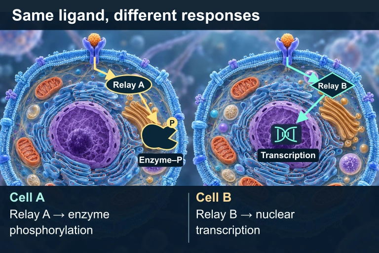 The same compatible ligand and receptor can produce different responses because the cells express different intracellular relays and effectors.
SECTION 4
Mechanisms of Transduction
Although signal transduction pathways vary enormously in their specific components, they rely on a surprisingly small toolkit of biochemical mechanisms. Understanding these mechanisms — phosphorylation cascades, second messenger systems, and protein conformational changes — is critical for interpreting experimental data on the AP exam.
Phosphorylation Cascades
The most common mechanism for relaying signals within the cytoplasm is the sequential activation of protein kinases — enzymes that transfer a phosphate group from ATP to a specific amino acid residue (typically serine, threonine, or tyrosine) on a target protein. Phosphorylation changes the target protein's shape and activity, often activating it so it can phosphorylate the next kinase in the chain. Each activated kinase can phosphorylate many copies of the next kinase, which is the molecular basis for signal amplification. The reverse process — removal of phosphate groups by protein phosphatases — inactivates the relay proteins and is crucial for signal termination. The balance between kinase and phosphatase activity determines the net signaling output at any given moment.
PHOSPHORYLATION REACTION
A kinase catalyzes the forward reaction; a phosphatase catalyzes the reverse (dephosphorylation), regenerating the inactive protein and releasing Pᵢ.
Second Messengers
Second messengers are small, non-protein, water-soluble molecules or ions that rapidly diffuse through the cytoplasm to propagate a signal. The term "second" distinguishes them from the "first" messenger — the extracellular ligand. The three most important second messengers for the AP exam are cyclic AMP (cAMP), calcium ions (Ca²⁺), and inositol trisphosphate (IP₃). Cyclic AMP is produced by adenylyl cyclase when it is activated by a G protein; cAMP then activates protein kinase A (PKA). IP₃, produced by the cleavage of PIP₂ by phospholipase C, binds to calcium channels on the endoplasmic reticulum, releasing Ca²⁺ into the cytosol. The released calcium can bind calmodulin, which in turn activates other enzymes. These second messengers allow a single receptor event to flood the cell interior, providing both speed and amplification.
cAMP PRODUCTION
Adenylyl cyclase is activated by the Gα subunit of a stimulatory G protein (Gₛ). cAMP is degraded by phosphodiesterase, which converts cAMP to AMP and terminates the signal.
G Protein-Coupled Receptor (GPCR) Pathway
GPCRs are the largest and most diverse family of membrane receptors. They share a common architecture: seven transmembrane α-helices with an extracellular ligand-binding domain and an intracellular domain that interacts with a heterotrimeric G protein (composed of Gα, Gβ, and Gγ subunits). Upon ligand binding, the receptor facilitates the exchange of GDP for GTP on the Gα subunit, causing Gα-GTP to dissociate and activate a downstream effector enzyme such as adenylyl cyclase or phospholipase C. The intrinsic GTPase activity of Gα hydrolyzes GTP back to GDP, turning the G protein off — an elegant built-in timer for signal termination.
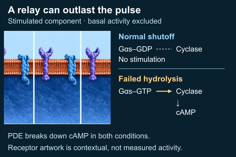 Inactive Gαs–GDP ceases to stimulate cyclase, while hydrolysis-defective Gαs–GTP can continue stimulation after the hormone pulse. PDE-mediated cAMP degradation continues in both. The selected signal-stimulated component excludes basal cyclase activity; repeated raster panels show contextual hardware, not measured states.
Receptor Tyrosine Kinase (RTK) Pathway
RTKs are single-pass transmembrane proteins that dimerize upon ligand binding, enabling their intracellular kinase domains to cross-phosphorylate each other on specific tyrosine residues. These phosphotyrosines serve as docking sites for various relay proteins containing SH2 domains, which then activate multiple downstream pathways simultaneously — including the Ras-MAPK pathway that regulates cell growth and division. Because a single activated RTK can recruit and activate many different relay proteins, RTKs are particularly well suited for triggering complex, multi-branched responses such as those controlling the cell cycle.
SECTION 5
Types of Cell Signaling & Receptor Classification
Cells communicate over distances ranging from fractions of a micrometer to meters, and the mode of signaling is classified according to this range. In addition, the location and type of receptor a cell uses determines which transduction pathway is engaged. The AP exam frequently asks you to distinguish among these categories, so a thorough understanding of each is essential.
| Feature | GPCR Pathway | RTK Pathway | Ligand-Gated Ion Channel |
|---|---|---|---|
| Signal speed | Moderate (seconds) | Moderate (seconds–minutes) | Very fast (milliseconds) |
| Amplification | High (via second messengers) | High (multi-branched cascades) | Low (direct ion flux) |
| Key second messenger | cAMP, IP₃, Ca²⁺ | Ras-GTP → MAPK cascade | Ions (Na⁺, K⁺, Ca²⁺) |
| Typical ligands | Epinephrine, glucagon, serotonin | Insulin, EGF, PDGF | Acetylcholine (nicotinic), GABA |
| Termination | GTPase on Gα; phosphodiesterase degrades cAMP | Phosphatases dephosphorylate RTK and targets | Channel closes when ligand dissociates |
Comparison of the three major membrane receptor-mediated signaling pathways
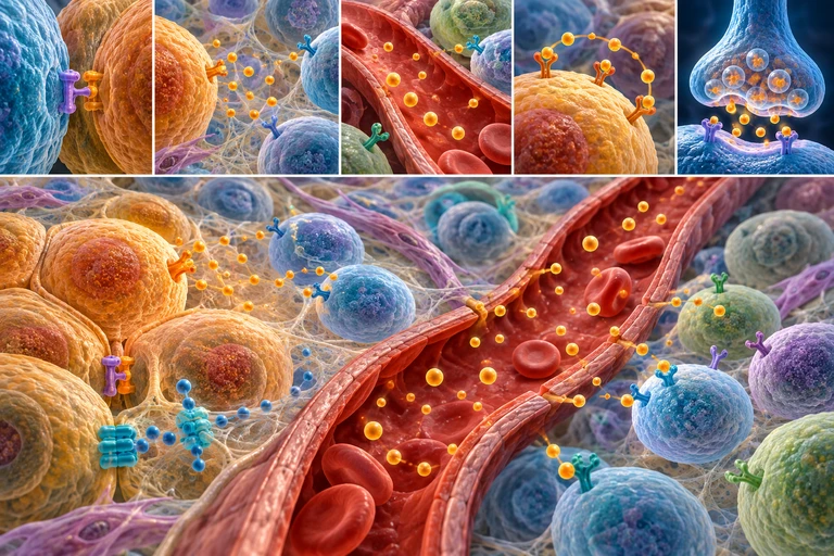 Cell-surface receptors connect extracellular ligand binding to intracellular responses. GPCRs, receptor tyrosine kinases and ligand-gated ion channels use distinct architectures.
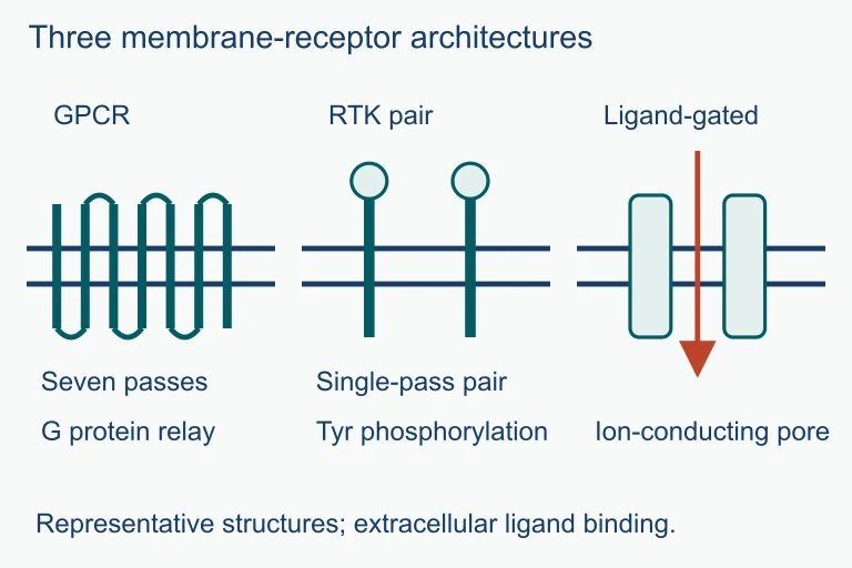 GPCRs relay signals through G proteins, receptor tyrosine kinases use phosphorylation, and ligand-gated channels open an ion-conducting pore.
SECTION 6
Worked Example: Epinephrine Signal Transduction
To solidify these concepts, let us trace the complete signal transduction pathway triggered when epinephrine (adrenaline) stimulates glycogen breakdown in a liver cell — the classic example studied by Sutherland.
Epinephrine → Glycogen Breakdown in a Liver Cell
- 1
Step 1 — Reception
Epinephrine, released from the adrenal medulla during a fight-or-flight response, circulates through the bloodstream and binds to a β-adrenergic receptor (a GPCR) on the surface of a hepatocyte. This binding is endocrine signaling because the signal molecule travels through the blood to a distant target.Receptor undergoes conformational change - 2
Step 2 — G Protein Activation
The activated receptor acts as a guanine nucleotide exchange factor (GEF), promoting the exchange of GDP for GTP on the Gα subunit of the associated stimulatory G protein (Gₛ). Gα-GTP dissociates from Gβγ and diffuses along the inner leaflet of the membrane.Gα-GTP is now active and mobile - 3
Step 3 — Adenylyl Cyclase Activation & cAMP Production
Gα-GTP binds and activates adenylyl cyclase, an integral membrane enzyme that converts ATP into cAMP. A single activated adenylyl cyclase can produce hundreds of cAMP molecules per second — this is the first major amplification step.Hundreds of cAMP molecules produced per receptor event - 4
Step 4 — Protein Kinase A (PKA) Activation
cAMP binds to the regulatory subunits of protein kinase A (PKA), releasing and activating its catalytic subunits. Each activated catalytic subunit can phosphorylate many copies of downstream target proteins — a second amplification step.PKA catalytic subunits phosphorylate phosphorylase kinase - 5
Step 5 — Phosphorylase Kinase → Glycogen Phosphorylase
Activated phosphorylase kinase phosphorylates glycogen phosphorylase, converting it to its active form (phosphorylase a). Each phosphorylase a molecule cleaves glucose-1-phosphate from glycogen at a high catalytic rate — a third amplification step.≈ 10⁸ glucose molecules released per initial epinephrine molecule - 6
Step 6 — Signal Termination
The signal is terminated at multiple levels: (1) the intrinsic GTPase activity of Gα hydrolyzes GTP → GDP, inactivating the G protein; (2) phosphodiesterase degrades cAMP → AMP; (3) protein phosphatases remove phosphate groups from phosphorylase kinase and glycogen phosphorylase, returning them to their inactive states.Signal fully terminated — system reset
AP Exam Tip
SECTION 7
Advantages & Vulnerabilities of Signal Transduction Systems
The multi-step architecture of signal transduction pathways confers remarkable advantages, but it also creates points of vulnerability. Understanding both sides is important for interpreting data on disrupted signaling — a frequent topic on the AP exam, particularly in the context of cancer and pharmacology.
| Advantage | Vulnerability |
|---|---|
| Amplification: A few ligand molecules generate millions of product molecules through cascaded enzymatic reactions. | Constitutively active kinases or G proteins (e.g., Ras mutations) bypass normal regulation, causing uncontrolled cell growth — a hallmark of many cancers. |
| Specificity: Different cell types express different receptors and relay proteins, so the same ligand can trigger distinct responses in different tissues. | Overexpression of receptors (e.g., HER2 in breast cancer) can make cells hypersensitive to growth signals that normal cells would ignore. |
| Integration: Cells can receive and process multiple signals simultaneously, integrating them through crosstalk between pathways to produce a coordinated response. | Inappropriate crosstalk can lead to pathway hijacking, where one pathway aberrantly activates another (e.g., inflammatory signaling promoting tumor survival). |
| Reversibility: Phosphatases and GTPases ensure rapid signal termination, allowing cells to respond dynamically to changing conditions. | Loss-of-function mutations in phosphatases or GTPases can prevent signal termination, causing sustained activation analogous to a stuck accelerator pedal. |
Strengths and vulnerabilities of multi-step signal transduction pathways
KEY TAKEAWAY
SECTION 8
Connection to the Cell Cycle & Advanced Topics
Signal transduction does not exist in isolation — it is the upstream regulatory mechanism that governs whether a cell enters, progresses through, or exits the cell cycle. Growth factors binding to RTKs activate the Ras-MAPK pathway, which ultimately upregulates cyclins — the proteins that drive cell cycle progression. Conversely, signals from neighboring cells or DNA damage checkpoints can activate pathways that halt division. The AP exam expects you to connect signal transduction directly to cell cycle regulation and to understand how defects in these pathways lead to cancer.
| Topic in This Lesson | Connection to Advanced Topic |
|---|---|
| Phosphorylation cascades | Cyclin-dependent kinases (CDKs) phosphorylate target proteins to advance the cell cycle through G₁, S, G₂, and M phases. |
| Ras-MAPK pathway (via RTKs) | Ras is one of the most frequently mutated proto-oncogenes in human cancers; a constitutively active Ras drives uncontrolled proliferation. |
| Signal termination & phosphatases | Tumor suppressors like p53 act as checkpoint monitors; loss of p53 function removes a key brake on the cell cycle, enabling cancer. |
| Receptor specificity | Targeted cancer therapies (e.g., imatinib for BCR-ABL, trastuzumab for HER2) exploit pathway-specific knowledge to selectively inhibit aberrant signals. |
| Second messengers (cAMP, Ca²⁺) | These molecules also regulate apoptosis (programmed cell death), immune responses, and neuronal plasticity — topics you will encounter in later AP Biology units. |
How signal transduction concepts connect to cell cycle regulation and cancer biology
As you progress through AP Biology, you will see signal transduction principles reappear in diverse contexts: immune cell activation, embryonic development, neuronal communication, and plant hormone responses. Mastering the general framework presented here — reception, transduction, response, amplification, and termination — equips you to analyze any specific pathway the exam may present, even ones you have never encountered before.
SECTION 9
Practice Problems
Question 1: CONCEPTUAL
- A. cAMP levels will decrease because the receptor can no longer bind its ligand.
- B. cAMP levels will remain at baseline because the drug targets the G protein, not adenylyl cyclase.
- C. cAMP levels will increase and remain elevated because adenylyl cyclase will be continuously activated.
- D. cAMP levels will initially increase but then rapidly decrease due to negative feedback from PKA.
Show answer (C)
Question 2: BASIC
- A. Epinephrine
- B. The β-adrenergic receptor
- C. Cyclic AMP (cAMP)
- D. Glycogen phosphorylase
Show answer (C)
Question 3: INTERMEDIATE
- A. The β-adrenergic receptor has a different structure in liver cells compared to cardiac muscle cells.
- B. Epinephrine is chemically modified as it travels from the liver to the heart.
- C. The two cell types express different sets of intracellular relay proteins and effector proteins downstream of the receptor.
- D. Cardiac muscle cells use cGMP instead of cAMP as the second messenger for epinephrine signaling.
Show answer (C)
Question 4: APPLIED
4 ptsShow answer
Question 5: CRITICAL THINKING
4 ptsShow answer
SUMMARY
Summary
Summary
Signal transduction is the process by which extracellular signals are converted into intracellular responses through a conserved three-stage framework: reception (ligand binds receptor), transduction (relay through phosphorylation cascades and second messengers such as cAMP, IP₃, and Ca²⁺), and response (altered gene expression, enzyme activity, or cell behavior). The three major membrane receptor families — GPCRs, receptor tyrosine kinases, and ligand-gated ion channels — each employ distinct mechanisms but share the principle that a small extracellular signal is amplified into a large intracellular effect.
Signal specificity depends on which receptors and downstream effectors a cell expresses, allowing the same ligand to produce different responses in different cell types. Signal termination — mediated by phosphatases, GTPase activity, and phosphodiesterase — ensures that responses are precise and reversible. Defects in signal transduction components, particularly oncogenes (constitutively active signaling proteins) and loss of tumor suppressors, are central to the molecular basis of cancer — a direct connection between signal transduction and cell cycle regulation that the AP exam heavily tests.