
SECTION 1
Historical Context & Motivation
One of the most profound questions in biology is deceptively simple: if every cell in a multicellular organism carries the same genome, how do cells become so different from one another? A neuron and a muscle fiber in the same human body share virtually identical DNA, yet they differ dramatically in morphology, protein composition, and function. The resolution of this paradox lies in differential gene expression—the selective activation and silencing of genes in different cell types. Understanding how organisms orchestrate gene expression across trillions of cells has been a central pursuit of molecular biology for over a century, and it remains one of the most actively studied topics in modern biomedical research.
1961 • Jacob & Monod: The Operon Model
1987 • Homeotic Genes & Hox Clusters
1998 • RNA Interference Discovered
2006 • Yamanaka & Induced Pluripotent Stem Cells
2012 • CRISPR-Cas9 Gene Editing
This historical trajectory reveals a recurring theme: the genome is not merely a static blueprint but a dynamically regulated information system. The central question that this lesson addresses is how eukaryotic cells deploy multiple layers of regulatory control—from chromatin remodeling to post-translational modification—to express only the subset of genes appropriate for a given cell type, developmental stage, or environmental condition.
SECTION 2
Core Principles of Differential Gene Expression
Cell specialization arises because different cell types transcribe different subsets of genes from an identical genome. This process of differential gene expression is governed by regulatory mechanisms operating at every stage of the pathway from DNA to functional protein. While every somatic cell in a human carries approximately 20,000–25,000 protein-coding genes, a typical differentiated cell expresses only a fraction of those genes at any given time. The following core principles form the foundation for understanding how cells achieve and maintain their specialized identities.
Genomic Equivalence
Hierarchical Regulation
Transcription Factors as Master Switches
Signal Transduction & Induction
Epigenetic Inheritance
KEY TAKEAWAY
SECTION 3
Levels of Gene Expression Regulation
The following diagram illustrates the major control points at which eukaryotic gene expression can be regulated, from the chromatin level through post-translational modification. Each level represents an opportunity for the cell to adjust the quantity, timing, and location of a specific protein, and collectively these regulatory layers produce the precise proteome that defines each cell type.
As shown in the diagram, the most consequential regulatory checkpoint is typically at the transcriptional level, where cells invest the most energy in controlling which genes are transcribed into mRNA. However, each downstream level—RNA processing, translation rate, and protein modification—provides additional fine-tuning. For example, alternative splicing at the post-transcriptional level allows a single gene to produce multiple distinct protein isoforms, dramatically expanding the proteome beyond the number of genes. The Drosophila Dscam gene can generate over 38,000 mRNA variants through alternative splicing—more protein variants than the total number of genes in the fly's genome.
SECTION 4
Mechanisms of Transcriptional Regulation
Because transcriptional regulation is the primary mechanism by which cells establish and maintain differentiated states, understanding the molecular details of this process is essential for the AP Biology exam. Eukaryotic transcription requires the assembly of a complex molecular apparatus at gene promoters, and the efficiency of this assembly is modulated by multiple classes of regulatory elements and proteins.
Promoters, Enhancers, and Silencers
Every eukaryotic gene has a promoter—a DNA sequence located immediately upstream of the transcription start site that serves as the binding platform for RNA polymerase II and general transcription factors. The promoter alone, however, typically supports only a basal (very low) rate of transcription. The cell-type-specific activation of genes depends heavily on enhancers—regulatory DNA sequences that can be located thousands of base pairs upstream or downstream of the gene they regulate, or even within introns. Enhancers function by binding specific activator proteins, which interact with the transcription initiation complex through DNA looping mediated by mediator proteins. Conversely, silencers are regulatory sequences that bind repressor proteins to inhibit transcription.
Epigenetic Modifications
The accessibility of DNA to transcription factors is largely controlled by epigenetic modifications. DNA methylation—the addition of methyl groups (—CH₃) to cytosine bases, particularly at CpG dinucleotides—is generally associated with gene silencing. Methylated DNA recruits proteins that condense chromatin, physically blocking transcription factor access. Histone acetylation, by contrast, loosens the association between histones and DNA by neutralizing the positive charges on histone tails, promoting an open chromatin conformation (euchromatin) that is accessible to the transcriptional machinery. Tightly packed, transcriptionally inactive chromatin is termed heterochromatin. Histone deacetylation and methylation of specific histone residues (e.g., H3K9me3) promote heterochromatin formation and gene silencing.
Non-Coding RNAs in Regulation
Post-transcriptional regulation adds another dimension to gene expression control. MicroRNAs (miRNAs) are small non-coding RNA molecules (approximately 22 nucleotides) that bind complementary sequences in the 3' untranslated region (UTR) of target mRNAs, leading to mRNA degradation or translational repression. A single miRNA species can regulate hundreds of target mRNAs, and a single mRNA can be targeted by multiple miRNAs. Small interfering RNAs (siRNAs) operate through a related mechanism and can also direct chromatin modifications. Together, these non-coding RNAs constitute a powerful regulatory network that fine-tunes gene expression during development and in response to environmental changes.
AP EXAM TIP
SECTION 5
From Stem Cells to Specialized Cells: Differentiation Pathways
Cell specialization during development follows a hierarchical branching pattern in which cells progressively restrict their developmental potential. A fertilized egg (zygote) is totipotent—capable of giving rise to every cell type in the organism plus extraembryonic tissues such as the placenta. As development proceeds, cells become pluripotent (able to form all cell types of the body but not extraembryonic tissues), then multipotent (restricted to a specific lineage, such as hematopoietic stem cells that produce all blood cell types), and finally terminally differentiated (committed to a single specialized function). At each branch point, inductive signals from neighboring cells and morphogen concentration gradients activate specific sets of transcription factors, which in turn establish new patterns of gene expression that are locked in by epigenetic modifications.
| Potency Level | Definition | Example |
|---|---|---|
| Totipotent | Can form all cell types plus extraembryonic tissues | Zygote; cells up to ~8-cell stage |
| Pluripotent | Can form all body cell types but not extraembryonic tissues | Inner cell mass (ICM) of blastocyst; iPSCs |
| Multipotent | Can form multiple cell types within a single lineage | Hematopoietic stem cells; neural stem cells |
| Unipotent | Can produce only one cell type; can self-renew | Muscle satellite cells; spermatogonial stem cells |
Potency levels during cell differentiation
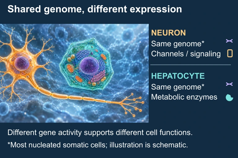 Neuron and hepatocyte share most of their genome but express different sets of genes. Illustration is schematic where scale or molecular structure is simplified.
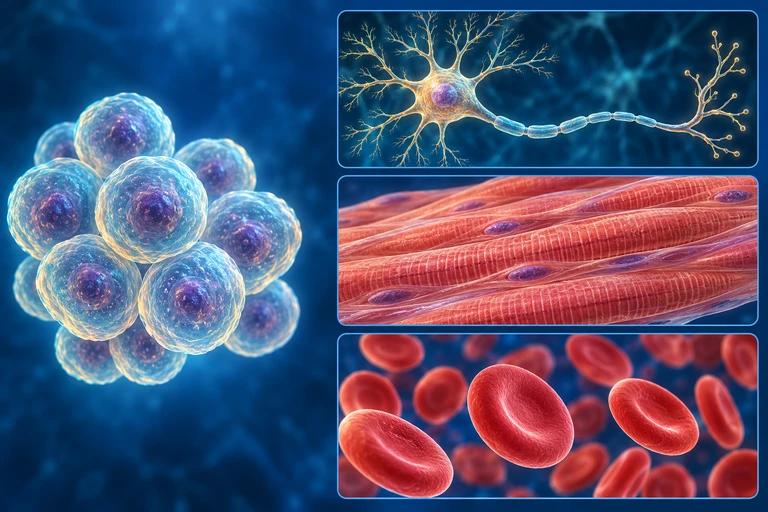 Differentiation restricts developmental potential through gene regulation. Reprogramming can restore pluripotency in nucleated somatic cells; mature mammalian red cells have lost their nuclei and cannot serve as the source cells in this route.
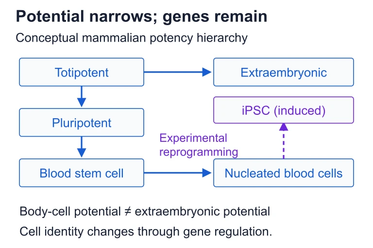 Differentiation restricts developmental potential through gene regulation. Reprogramming can restore pluripotency in nucleated somatic cells; mature mammalian red cells have lost their nuclei and cannot serve as the source cells in this route.
SECTION 6
Worked Example: The lac Operon as a Model of Gene Regulation
Although the lac operon is a prokaryotic system, it remains one of the clearest models for understanding the logic of gene regulation and is frequently tested on the AP Biology exam. The principles of inducible gene expression it demonstrates—signal-dependent activation, repressor proteins, and positive regulation—are conceptually analogous to mechanisms operating in eukaryotic cells.
Predicting lac Operon Expression Under Different Conditions
- 1
Step 1 — Identify the Regulatory Components
The lac operon consists of three structural genes (lacZ, lacY, lacA), a promoter, an operator, and the separately transcribed lacI regulatory gene that encodes the lac repressor protein. Two signals matter: the presence of lactose (inducer) and the level of glucose (detected via cAMP/CAP). - 2
Step 2 — Assess Condition: Glucose Present, Lactose Absent
When glucose is available and lactose is absent, the cell has no need to express lactose-metabolizing enzymes. The lac repressor binds the operator, physically blocking RNA polymerase. Additionally, glucose suppresses cAMP levels, so the CAP activator protein cannot bind the promoter region. Result: operon OFF.Transcription: OFF (repressor bound, no CAP activation) - 3
Step 3 — Assess Condition: Glucose Absent, Lactose Present
Without glucose, cAMP levels rise, and the cAMP–CAP complex binds upstream of the promoter, enhancing RNA polymerase binding. Simultaneously, allolactose (derived from lactose) binds the lac repressor, causing a conformational change that releases it from the operator. Both negative regulation (repressor removal) and positive regulation (CAP activation) favor maximal transcription.Transcription: MAXIMAL (repressor released + CAP bound) - 4
Step 4 — Apply to Eukaryotic Analogy
In eukaryotic cells, the logic is analogous but far more complex. Instead of a single repressor/operator system, a eukaryotic gene may have dozens of enhancers, each bound by different transcription factors. The combinatorial presence of these factors—determined by the cell's developmental history and current signaling environment—determines whether a gene is actively transcribed, providing the molecular basis for cell specialization. - 5
Step 5 — Summarize the Regulatory Logic
Gene expression is controlled by the interplay of positive regulators (activators, enhancers, CAP) and negative regulators (repressors, silencers, methylation). Whether in prokaryotes or eukaryotes, gene regulation allows cells to express the right genes at the right time in the right amounts.Core principle: Differential gene expression = combinatorial control by activators + repressors + epigenetic state
SECTION 7
Prokaryotic vs. Eukaryotic Gene Regulation
While the fundamental logic of gene regulation—using regulatory proteins to control transcription in response to signals—is conserved across all domains of life, prokaryotic and eukaryotic organisms differ substantially in the complexity and mechanisms of their regulatory systems. Understanding these differences is essential for the AP Biology exam, which frequently presents comparative questions.
| Feature | Prokaryotic Regulation | Eukaryotic Regulation |
|---|---|---|
| Gene organization | Genes grouped in operons; polycistronic mRNA | Individual genes with own promoters; monocistronic mRNA |
| Chromatin structure | No histones (nucleoid-associated proteins instead); DNA is accessible | DNA wrapped around histones; chromatin remodeling required for access |
| Primary control point | Transcription initiation (repressors/activators at operator) | Multiple levels; transcription initiation is primary but epigenetic and post-transcriptional layers are extensive |
| Enhancers | Rare; CAP site is a simple positive element | Abundant; can be >1 Mb from gene; act through DNA looping |
| RNA processing | Minimal; no introns, no splicing, no 5' cap or poly-A tail | Extensive; 5' capping, 3' polyadenylation, intron splicing, alternative splicing |
| Coupling of transcription & translation | Simultaneous; ribosomes translate mRNA as it is being transcribed | Separated by nuclear envelope; mRNA must be exported to cytoplasm before translation |
Comparison of prokaryotic and eukaryotic gene regulation
KEY TAKEAWAY
SECTION 8
Connections to Cancer, Development & Biotechnology
Disruptions in gene expression regulation are not merely academic curiosities—they underlie some of the most significant phenomena in biology and medicine. When the regulatory mechanisms that maintain cell specialization go awry, the consequences can be catastrophic, as seen in cancer. Conversely, deliberate manipulation of gene expression has opened transformative possibilities in biotechnology and regenerative medicine.
| Topic | Connection to Gene Expression Regulation |
|---|---|
| Cancer | Mutations in proto-oncogenes (gain-of-function → oncogenes) or tumor suppressor genes (loss-of-function) disrupt normal growth signaling. Epigenetic silencing of tumor suppressors via promoter hypermethylation is also common. Cancer cells essentially "de-differentiate," losing specialized functions and reverting to uncontrolled proliferation. |
| Embryonic Development | Morphogen gradients (e.g., bicoid in Drosophila) establish positional information that activates cascades of transcription factors. Hox genes specify anterior-posterior body axis identity. Mutations in these master regulators cause dramatic homeotic transformations—legs growing where antennae should be. |
| iPSC Technology | Yamanaka's four factors (Oct4, Sox2, Klf4, c-Myc) are transcription factors that reset the epigenetic landscape of differentiated cells, restoring pluripotency. This demonstrates that differentiation is maintained by gene expression patterns, not by irreversible DNA loss. |
| CRISPR & Gene Therapy | CRISPR-Cas9 can edit genes, but modified versions (CRISPRa, CRISPRi) can activate or silence genes without changing the DNA sequence—essentially reprogramming gene expression at will. These tools hold promise for correcting gene regulation defects in genetic diseases. |
| X-Inactivation | In female mammals, one X chromosome per cell is epigenetically silenced by Xist long non-coding RNA and heterochromatin formation. This dosage compensation mechanism illustrates how epigenetic regulation can silence an entire chromosome while leaving the other active, even though both carry the same sequences. |
Advanced connections between gene expression regulation and key biological/medical topics
For the AP Biology exam, be prepared to explain how disruptions in gene regulation—whether caused by mutations, epigenetic changes, or environmental exposures—can lead to disease states. Additionally, understand that biotechnological interventions like CRISPR and iPSC generation work precisely because cell identity is determined by reversible gene expression patterns rather than permanent changes to the DNA sequence. These connections between gene regulation, development, disease, and technology are high-yield topics that integrate multiple units of the AP Biology curriculum.
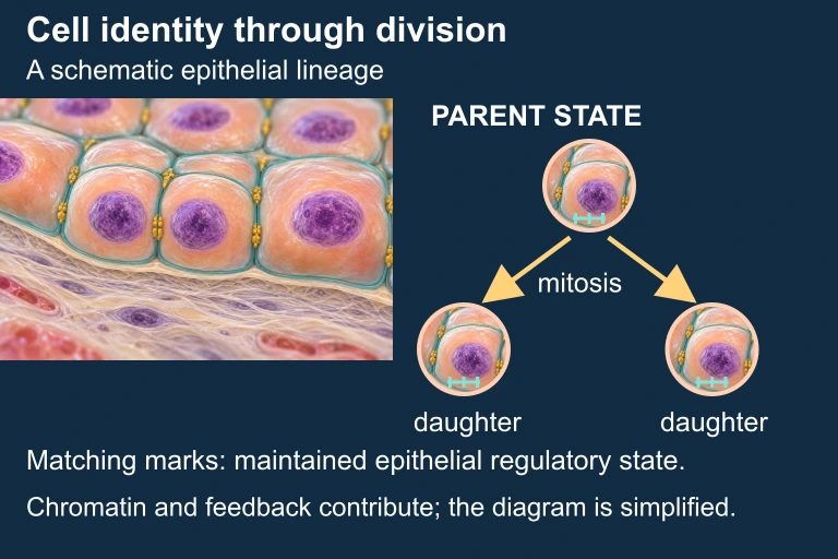 Cell division can preserve a differentiated regulatory state without changing the DNA sequence. Illustration is schematic where scale or molecular structure is simplified.
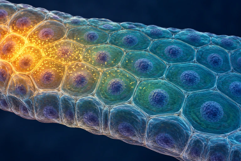 Cells at different positions receive different morphogen concentrations. At matched exposure time and competence, concentration thresholds can produce distinct response domains; other signals also influence cell fate.
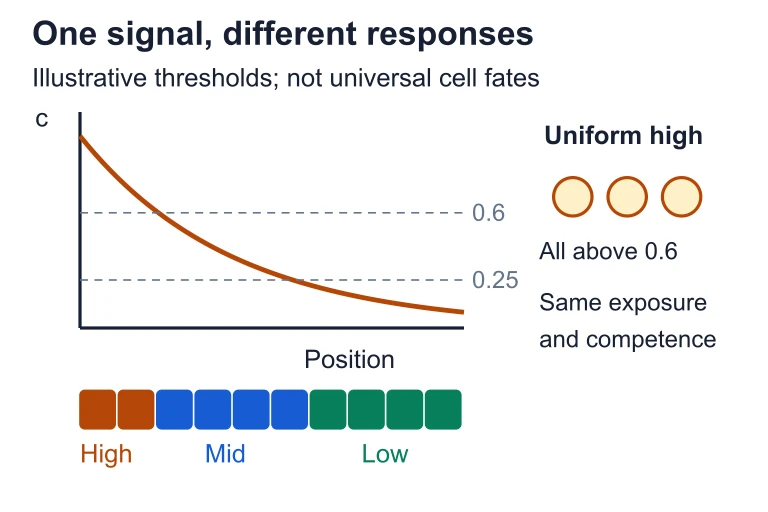 Cells at different positions receive different morphogen concentrations. At matched exposure time and competence, concentration thresholds can produce distinct response domains; other signals also influence cell fate.
SECTION 9
Practice Problems
Question 1: CONCEPTUAL
- A. Neurons undergo somatic mutations that delete liver-specific genes during development.
- B. Different transcription factors and epigenetic marks in each cell type activate different subsets of genes.
- C. Neurons and hepatocytes have different numbers of chromosomes due to selective chromosome loss.
- D. Each cell type uses a different genetic code to translate mRNA into different proteins.
Show answer (B)
Question 2: BASIC
- A. Genes that were previously silenced by methylation will become activated.
- B. All transcription in the cell will cease because RNA polymerase requires methylation to function.
- C. Histone acetylation levels will decrease, leading to more condensed chromatin.
- D. The DNA sequence of CpG islands will be altered, causing permanent mutations.
Show answer (A)
Question 3: INTERMEDIATE
- A. Cells throughout the neural tube will differentiate into floor plate neurons.
- B. Cells throughout the neural tube will differentiate into interneurons because high Shh is toxic.
- C. Normal patterning will occur because cells can detect absolute Shh concentration independently of the gradient.
- D. Cells will fail to differentiate entirely and will remain as undifferentiated progenitor cells.
Show answer (A)
Question 4: APPLIED
4 ptsShow answer
Question 5: CRITICAL THINKING
4 ptsShow answer
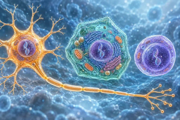 Nearly identical genomes support different expression profiles. Gene W is broadly expressed in these cells, while X, Y and Z are enriched in neurons, hepatocytes and embryonic stem cells, respectively.
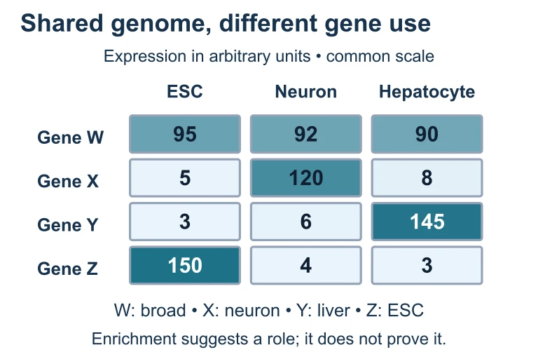 Nearly identical genomes support different expression profiles. Gene W is broadly expressed in these cells, while X, Y and Z are enriched in neurons, hepatocytes and embryonic stem cells, respectively.
SUMMARY
Gene Expression and Cell Specialization — Key Concepts
Summary
All somatic cells in a multicellular organism share the same genome, and cell specialization arises through differential gene expression—the selective activation and silencing of specific gene subsets. This regulation operates at multiple hierarchical levels: epigenetic control (DNA methylation and histone modifications that alter chromatin accessibility), transcriptional regulation (transcription factors binding promoters, enhancers, and silencers), post-transcriptional regulation (alternative splicing, miRNA-mediated mRNA degradation), translational control, and post-translational modifications (phosphorylation, ubiquitination, proteolytic cleavage).
During development, cells progress from totipotent to pluripotent to multipotent to terminally differentiated states, guided by inductive signals such as morphogen gradients and cell-cell contacts that activate specific transcription factor cascades. Disruptions in gene regulation underlie diseases such as cancer, while deliberate reprogramming through factors like the Yamanaka factors can reverse differentiation, confirming that cell identity is encoded not in DNA sequence changes but in the dynamic, epigenetically maintained landscape of gene expression.