

SECTION 1
Historical Context & Motivation
The realization that living organisms rely on specific catalytic molecules—enzymes—to drive biochemical reactions was one of the great achievements of nineteenth- and twentieth-century biology. Early fermentation studies revealed that yeast extracts could convert sugar into alcohol even outside of living cells, but the question of why these catalysts were so sensitive to heat, acidity, and other environmental conditions remained unanswered for decades. Understanding how environmental variables alter enzyme activity is not merely an academic exercise: it explains phenomena as diverse as why fevers can be dangerous, why stomach acid aids digestion, and why organisms living in hot springs require specialized proteins. Today, this knowledge underpins industrial biotechnology, medicine, and ecological physiology, making it a cornerstone of the AP Biology curriculum.
1833 • Payen & Persoz Isolate Diastase
1897 • Buchner's Cell-Free Fermentation
1913 • Michaelis–Menten Kinetics
1926 • Sumner Crystallizes Urease
1960s–1970s • Thermophilic Enzymes Discovered
These milestones reveal a recurring theme: an enzyme's catalytic efficiency is inseparable from its molecular environment. The central question that frames this lesson is straightforward yet profound—how do temperature, pH, salinity, and other environmental factors alter the three-dimensional structure of an enzyme, and why does that structural change affect catalytic activity? Answering this question requires integrating concepts of protein folding, thermodynamics, and evolutionary adaptation.
SECTION 2
Core Principles & Definitions
Before exploring how environmental factors modulate enzyme function, it is essential to ground yourself in several core principles. Enzymes are biological catalysts—typically proteins, though some RNA molecules (ribozymes) also catalyze reactions—that lower the activation energy (Ea) of a reaction without being consumed. Their catalytic power depends on the precise geometry of the active site, a region shaped by the enzyme's overall three-dimensional conformation. Any environmental change that disrupts this conformation will consequently alter catalytic efficiency—sometimes enhancing it, often diminishing it, and in extreme cases abolishing it entirely through denaturation.
Activation Energy & Catalysis
Active Site & Induced Fit
Optimal Conditions
Denaturation
Cofactors & Coenzymes
KEY TAKEAWAY
SECTION 3
Visualizing Temperature & pH Effects
The relationship between environmental variables and enzyme activity is best understood graphically. The following diagram illustrates two classic curves: the bell-shaped temperature–activity curve and the pH–activity curve. Notice that both curves are asymmetric: the decline past the optimum is often steeper than the ascent, reflecting the irreversible nature of denaturation at extreme conditions.
Several important patterns emerge from these curves. First, both temperature and pH curves are roughly bell-shaped, confirming that enzyme activity is maximal at a specific optimum and declines on either side. Second, the shape of each curve varies by enzyme: thermophilic enzymes found in hot-spring archaea have temperature optima above 80 °C, while psychrophilic enzymes in polar fish operate optimally near 10 °C. Third, the width of the bell reflects the enzyme's tolerance range—some enzymes are broadly tolerant, while others function within a very narrow window. These differences are evolutionarily significant: natural selection molds enzyme structure to match the organism's habitat.
SECTION 4
Molecular Mechanisms of Environmental Effects
Temperature Effects: Kinetic Energy vs. Structural Stability
Temperature influences enzyme activity through two competing mechanisms. On one hand, increasing temperature raises the average kinetic energy of both enzyme and substrate molecules, increasing the frequency and energy of molecular collisions and thus accelerating the rate at which substrate enters the active site. This relationship is described by the Arrhenius equation, which predicts that reaction rate roughly doubles for every 10 °C increase (the Q10 rule). On the other hand, higher temperatures also increase molecular vibrations that disrupt the weak non-covalent interactions—hydrogen bonds, van der Waals forces, ionic bonds, and hydrophobic interactions—that stabilize the enzyme's tertiary structure. When these bonds break faster than they reform, the enzyme undergoes thermal denaturation, irreversibly losing its active-site geometry.
Q₁₀ TEMPERATURE COEFFICIENT
Where R₁ and R₂ are reaction rates at temperatures T₁ and T₂ (in °C). A Q10 of ≈ 2 means the rate doubles with each 10 °C increase, which holds only below the denaturation threshold.
pH Effects: Ionization of Active-Site Residues
The pH of the surrounding solution determines the protonation state of ionizable amino acid side chains (e.g., histidine, aspartate, glutamate, lysine) within the active site. Because substrate binding and catalysis often depend on specific electrostatic interactions between charged residues and the substrate, even a small shift in pH can alter these charges and reduce catalytic efficiency. At pH values far from the optimum, widespread changes in charge distribution destabilize the entire protein, causing acid or alkaline denaturation. It is important to recognize that optimal pH is not universal—pepsin (stomach protease) functions optimally at pH 2 because its active-site aspartate residues are properly protonated at that pH, while trypsin (intestinal protease) requires pH 8 for the correct ionization state of its catalytic triad.
Salt Concentration & Ionic Strength
Ionic strength affects enzyme function by screening electrostatic interactions between charged residues. Moderate salt concentrations can stabilize an enzyme's native conformation by shielding repulsive charges, but excessive salt disrupts the hydration shell around the protein and can cause salting out—precipitation of the protein from solution. Halophilic organisms (salt-loving archaea) have evolved enzymes with excess negatively charged surface residues that require high salt to neutralize charge repulsion and maintain proper folding, illustrating the coevolution of enzyme structure and habitat.
Substrate Concentration
While substrate concentration is not strictly an 'environmental' variable in the same sense as temperature or pH, it is a critical context that the AP exam expects you to understand. At low [S], the reaction rate increases almost linearly with substrate availability because many active sites are unoccupied. As [S] rises, the rate increase slows, and eventually the enzyme reaches Vmax—the maximum velocity at which all active sites are saturated. This saturation behavior is captured by the Michaelis–Menten equation.
MICHAELIS–MENTEN EQUATION
Where v = reaction velocity, Vmax = maximum velocity at substrate saturation, [S] = substrate concentration, and Km = Michaelis constant (the [S] at which v = ½Vmax). Environmental factors like temperature and pH can alter both Vmax and Km.
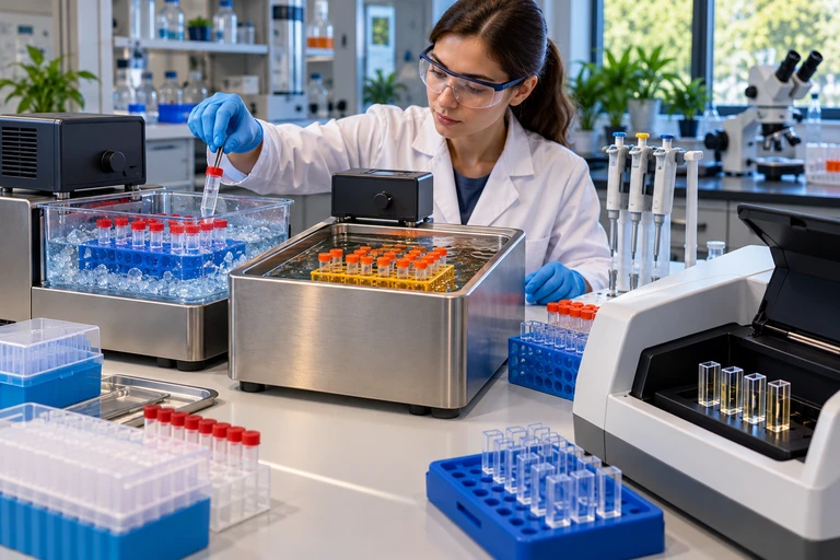 To distinguish prior temperature exposure from immediate reaction conditions, return all enzyme treatments to the same assay temperature and pH. Recovery after cooling differs from persistent activity loss after heating.
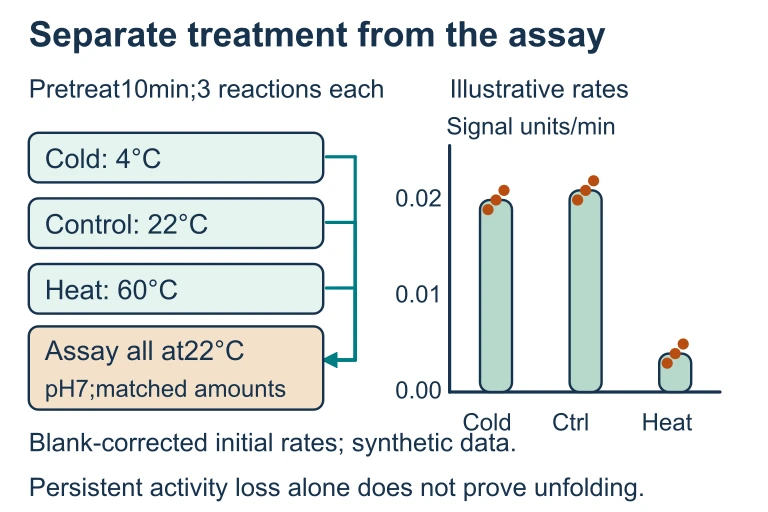 To distinguish prior temperature exposure from immediate reaction conditions, return all enzyme treatments to the same assay temperature and pH. Recovery after cooling differs from persistent activity loss after heating.
SECTION 5
Comparing Environmental Factors in Detail
The following diagram provides a comprehensive comparison of how four major environmental factors affect the same hypothetical enzyme. Studying these curves side by side reinforces a critical concept: each factor has an optimum, and deviations from that optimum reduce activity through distinct but overlapping molecular mechanisms.
| Factor | Molecular Mechanism | Effect at Optimum | Effect at Extreme |
|---|---|---|---|
| Temperature | Increases kinetic energy of molecules, raising collision frequency; simultaneously increases molecular vibrations that disrupt non-covalent bonds | Maximal Vmax; ideal balance of collision energy and structural integrity | Thermal denaturation; irreversible unfolding of tertiary/quaternary structure |
| pH | Alters protonation state of ionizable amino acid side chains (His, Asp, Glu, Lys, Arg), affecting electrostatic interactions and active-site charge distribution | Correct ionization of catalytic residues; optimal substrate binding and transition-state stabilization | Acid or alkaline denaturation; widespread disruption of ionic bonds throughout protein |
| Salt / Ionic Strength | Ions screen electrostatic interactions between charged residues; high concentrations compete for the protein's hydration shell | Stabilized conformation via charge screening; proper folding maintained | Salting out (precipitation); loss of hydration shell; aggregation |
| Substrate [S] | Governs active-site occupancy; more substrate increases the probability of productive enzyme–substrate encounters | At [S] >> Km, all active sites saturated → Vmax | Substrate inhibition can occur in some enzymes at very high [S], though this is not universal |
Summary comparison of four major environmental factors affecting enzyme function
SECTION 6
Worked Example: Predicting Enzyme Behavior
The following worked example walks through a data-interpretation scenario typical of AP Biology free-response questions. Practice reasoning through each step, as this type of problem demands both conceptual understanding and the ability to connect environmental conditions to molecular-level changes.
Predicting Catalase Activity Under Changing Conditions
- 1
Step 1 — Identify the Enzyme and Its Context
Catalase decomposes hydrogen peroxide (2H₂O₂ → 2H₂O + O₂) in nearly all aerobic organisms. In humans, catalase operates in peroxisomes and has an optimal temperature of approximately 37 °C and an optimal pH near 7.0. - 2
Step 2 — Analyze the Scenario
A student measures O₂ production (a proxy for reaction rate) from beef liver catalase at 10 °C, 25 °C, 37 °C, 50 °C, and 70 °C, each at pH 7.0. She obtains the highest O₂ production at 37 °C, moderate production at 25 °C and 50 °C, low production at 10 °C, and nearly zero production at 70 °C. - 3
Step 3 — Explain the Low-Temperature Result
At 10 °C, molecules have lower kinetic energy, so collisions between catalase and H₂O₂ are less frequent and less energetic. Fewer substrate molecules have sufficient energy to reach the transition state, resulting in a slower reaction rate. Importantly, the enzyme is not denatured—it is simply less active.Low temperature → reduced kinetic energy → slower rate (enzyme intact) - 4
Step 4 — Explain the High-Temperature Result
At 70 °C, thermal energy is sufficient to disrupt the hydrogen bonds, ionic interactions, and hydrophobic contacts that stabilize catalase's tertiary structure. The active site loses its complementary shape, and substrate can no longer bind effectively. Because denaturation of catalase is essentially irreversible under these conditions (the protein aggregates), cooling the sample back to 37 °C will not restore activity.70 °C → thermal denaturation → irreversible loss of active-site geometry - 5
Step 5 — Predict pH Shift Effect
If the experiment were repeated at pH 2 instead of pH 7, catalase activity would be dramatically reduced even at 37 °C. At pH 2, the high concentration of H⁺ ions protonates negatively charged side chains (Asp, Glu), disrupting ionic bonds and altering the charge distribution within the active site. The enzyme would denature under strongly acidic conditions, demonstrating that temperature and pH are independent variables that both must be near their optima for maximal activity.pH 2 at 37 °C → acid denaturation → near-zero activity despite optimal temperature
SECTION 7
Evolutionary Adaptations & Limitations
A fascinating consequence of the environmental sensitivity of enzymes is that organisms living in extreme habitats have evolved enzyme variants—extremozymes—with shifted optima. These adaptations underscore a broader evolutionary principle: natural selection acts on protein structure to match catalytic function to ecological context. However, every adaptation involves trade-offs. Thermostable enzymes, for instance, tend to be more rigid at mesophilic temperatures, reducing their catalytic rate under 'normal' conditions. The table below compares enzyme adaptations across several ecological niches.
| Organism Type | Habitat | Enzyme Adaptation | Trade-Off |
|---|---|---|---|
| Thermophiles | Hot springs, hydrothermal vents (60–120 °C) | More disulfide bonds, more ionic bridges, tightly packed hydrophobic cores increase structural stability at high temperatures | Reduced flexibility (and therefore reduced catalytic efficiency) at lower temperatures |
| Psychrophiles | Polar oceans, glacial ice (−2 to 10 °C) | Fewer stabilizing bonds, more glycine residues (greater backbone flexibility), larger active-site cavities enhance catalysis at low temperatures | Extremely heat-labile; denaturation occurs at moderate temperatures (≈ 30–40 °C) |
| Acidophiles | Acid mine drainage, volcanic soils (pH 1–3) | Excess negative surface charges; intracellular pH maintained near neutral by proton pumps, protecting cytoplasmic enzymes | Enzymes denature rapidly at neutral or alkaline pH |
| Halophiles | Salt lakes, salt flats (2–5 M NaCl) | Excess acidic residues on protein surface require high salt to neutralize charge repulsion; increased hydration by salt ions | Enzymes unfold and lose activity in low-salt environments |
Enzyme adaptations across extremophilic organisms
KEY TAKEAWAY
SECTION 8
Connections to Advanced Topics
The principles you have learned about environmental impacts on enzyme function connect directly to several advanced topics in biology and biochemistry. Understanding these connections will help you integrate knowledge across AP Biology units and prepare for synthesis-style exam questions. In particular, the relationship between enzyme behavior and homeostasis (Unit 8) is a high-value connection: organisms invest substantial energy in maintaining stable internal conditions precisely because their enzymes are so environmentally sensitive.
| Concept in This Lesson | Advanced / Cross-Unit Connection |
|---|---|
| Optimal temperature for human enzymes ≈ 37 °C | Thermoregulation (Unit 8): Endothermy maintains body temperature in the narrow range where metabolic enzymes function optimally; fever represents a controlled departure that can impair pathogen enzymes while still within the tolerance range of host enzymes |
| pH sensitivity of active-site residues | Buffer systems (Chemistry connection): Bicarbonate buffer in blood maintains plasma pH near 7.4; respiratory and renal compensations protect enzyme function during metabolic acid–base disturbances |
| Denaturation as irreversible unfolding | Chaperone proteins (molecular chaperones like Hsp70) assist in protein refolding under stress; heat-shock response is a gene regulation topic (Unit 6) triggered when temperatures approach the denaturation threshold |
| Extremozyme evolution | Natural selection and adaptation (Unit 7): Enzyme gene sequences in extremophiles show evidence of positive selection at structurally critical residue positions; molecular phylogenetics can trace the evolution of thermostability |
| Michaelis–Menten kinetics | Allosteric regulation and feedback inhibition (Unit 3): Environmental conditions that alter Km or Vmax interact with competitive and noncompetitive inhibitors to fine-tune metabolic flux |
Cross-unit connections linking enzyme environmental sensitivity to other AP Biology topics
As you advance beyond AP Biology into college biochemistry, you will encounter more sophisticated treatments of these topics, including transition-state theory (which rigorously derives the Arrhenius equation from statistical mechanics), protein folding energy landscapes (funnel models of how polypeptides navigate conformational space), and directed evolution (laboratory techniques that mimic natural selection to engineer enzymes with novel environmental tolerances). The conceptual foundation you build now—environment alters structure, structure determines function—will serve as the scaffold for all of these advanced ideas.
SECTION 9
Practice Problems
Question 1: CONCEPTUAL
- A. At 60 °C, substrate molecules move too fast to bind the active site, so the reaction cannot proceed.
- B. At 60 °C, the activation energy of the reaction increases beyond what the enzyme can lower.
- C. At 60 °C, thermal energy disrupts non-covalent bonds maintaining the enzyme's tertiary structure, denaturing the active site so that substrate can no longer bind effectively.
- D. At 60 °C, the enzyme is consumed more rapidly in the reaction, so none remains to catalyze further substrate conversion.
Show answer (C)
Question 2: BASIC
- A. Pepsin and trypsin have identical amino acid sequences but fold differently depending on ambient pH.
- B. Pepsin's active-site residues are properly ionized for catalysis at low pH, while trypsin's active-site residues require the ionization states present at alkaline pH.
- C. Trypsin requires a cofactor that is only stable at pH 8, whereas pepsin's cofactor is stable at pH 2.
- D. The stomach provides more thermal energy than the small intestine, and pepsin uses this extra energy in place of a higher pH.
Show answer (B)
Question 3: INTERMEDIATE
- A. Number of glycine residues in flexible loop regions
- B. Number of disulfide bonds and salt bridges
- C. Overall molecular weight of the enzyme
- D. Number of active sites per polypeptide chain
Show answer (B)
Question 4: APPLIED
4 ptsShow answer
Question 5: CRITICAL THINKING
4 ptsShow answer
SUMMARY
Lesson Summary
Summary
Enzymes are biological catalysts whose function depends entirely on maintaining the correct three-dimensional conformation of their active site. Environmental factors—temperature, pH, salt concentration, and substrate concentration—modulate enzyme activity by affecting this conformation. Each enzyme exhibits an optimal temperature and optimal pH at which catalytic rate is maximal. Below the optimum, reduced kinetic energy slows molecular collisions; above the optimum, thermal energy or altered ionization states disrupt non-covalent bonds and cause denaturation—an often irreversible loss of functional shape.
Organisms in extreme environments have evolved extremozymes with shifted optima—thermophilic enzymes with extra disulfide bonds and salt bridges for heat resistance, and psychrophilic enzymes with increased glycine content and larger active-site cavities for cold flexibility. The unifying principle is that structure determines function: any factor that alters protein structure will alter enzyme activity. For the AP exam, remember to connect environmental impacts on enzymes to broader themes including homeostasis, natural selection, and the Michaelis–Menten model of enzyme kinetics.