
SECTION 1
Historical Context & Motivation
The discovery that all living organisms are composed of cells ranks among the most transformative insights in the history of biology. Before the invention of the microscope, scholars could only speculate about the fundamental units of life, relying on philosophical frameworks rather than empirical evidence. The gradual refinement of optical instruments from the seventeenth century onward opened a previously invisible world, revealing that tissues, organs, and entire organisms are built from discrete structural units. This realization culminated in the cell theory, a unifying principle that remains foundational to modern biology, medicine, and biotechnology.
1665 • Robert Hooke Coins "Cell"
1674 • Van Leeuwenhoek Observes Living Cells
1838–1839 • Schleiden & Schwann: Cell Theory
1855 • Virchow: Omnis Cellula e Cellula
1950s–1970s • Electron Microscopy & Endosymbiotic Theory
The central question that cell biology addresses is deceptively simple: How does the internal organization of a cell give rise to the emergent properties of life? From energy conversion to gene expression, every biological process depends on the spatial arrangement of membranes, enzymes, and genetic material within cells. Understanding cell structure is therefore prerequisite to understanding virtually every topic on the AP Biology exam — from cellular energetics to signal transduction to heredity.
SECTION 2
Core Principles of Cell Biology
Several foundational ideas underpin our understanding of cell structure and function. The modern cell theory extends the classical formulation of Schleiden, Schwann, and Virchow by incorporating insights from molecular biology and evolutionary theory. These principles organize the vast diversity of cell types into a coherent framework and explain why cells share certain universal features despite their morphological and functional specializations.
All Organisms Are Composed of Cells
Cells Arise from Pre-Existing Cells
Compartmentalization Enables Complexity
Surface-Area-to-Volume Ratio Constrains Cell Size
Endosymbiosis Explains Organelle Origin
SECTION 3
Visual Explanation — The Eukaryotic Cell
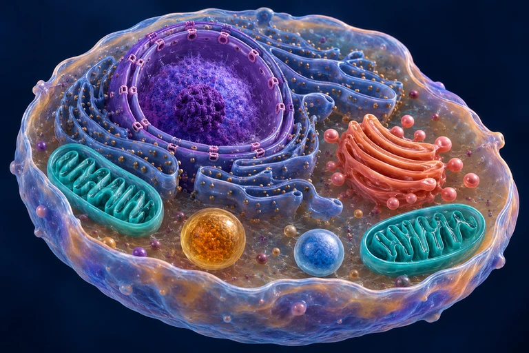
The diagram above illustrates the key architectural features of a typical animal cell. Notice that the cell is bounded by a single plasma membrane composed of a phospholipid bilayer studded with proteins, which regulates the passage of ions, nutrients, and signaling molecules. Within the cytoplasm, the endomembrane system — comprising the nuclear envelope, endoplasmic reticulum, Golgi apparatus, lysosomes, and vesicles — forms a functionally interconnected network that synthesizes, modifies, and distributes macromolecules. The spatial separation of these compartments ensures that enzymes with conflicting requirements (e.g., lysosomal hydrolases active at pH ≈ 5 versus cytoplasmic enzymes optimal near pH ≈ 7.2) do not interfere with one another, a critical aspect of cellular homeostasis.
 Membranes separate cytosol from organelle lumens and maintain distinct chemical environments. The nuclear outer membrane connects with the endoplasmic reticulum; mitochondrial and peroxisomal protein import follows routes distinct from ER–Golgi vesicle traffic.
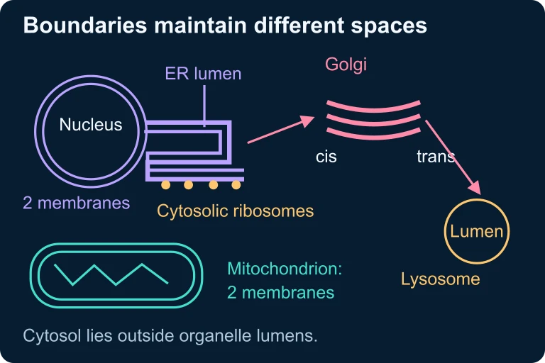 Membranes separate cytosol from organelle lumens and maintain distinct chemical environments. The nuclear outer membrane connects with the endoplasmic reticulum; mitochondrial and peroxisomal protein import follows routes distinct from ER–Golgi vesicle traffic.
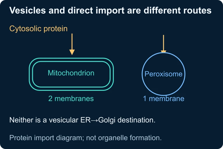 Mitochondrial and peroxisomal proteins can enter from the cytosol through direct-import routes distinct from ER–Golgi vesicle transport.
SECTION 4
How It Works — Membrane Structure and Transport
The plasma membrane is far more than a passive boundary; it is a dynamic, selectively permeable structure described by the fluid mosaic model proposed by Singer and Nicolson in 1972. In this model, a bilayer of phospholipids forms the structural foundation, with the hydrophilic heads oriented outward toward aqueous environments and the hydrophobic fatty acid tails oriented inward, creating a nonpolar interior that acts as a permeability barrier. Integral and peripheral membrane proteins are embedded within or associated with this bilayer, performing functions that include transport, enzymatic activity, signal transduction, cell–cell recognition, intercellular joining, and cytoskeleton attachment.
Transport Across the Membrane
Substances cross the plasma membrane via several mechanisms, broadly categorized as passive transport (no energy input required, movement down the concentration gradient) and active transport (requires ATP or another energy source, movement against the gradient). Passive transport includes simple diffusion of small nonpolar molecules like O₂ and CO₂ directly through the bilayer, osmosis (diffusion of water across a selectively permeable membrane), and facilitated diffusion through channel proteins or carrier proteins. Active transport includes primary active transport via ATP-hydrolyzing pumps such as the Na⁺/K⁺-ATPase, and secondary active transport (cotransport), which couples the movement of one solute down its gradient with the movement of another solute against its gradient.
SURFACE-AREA-TO-VOLUME RATIO
For a spherical cell, SA = surface area, V = volume, r = radius. As r increases, the ratio decreases, limiting the cell's ability to exchange materials efficiently. This inverse relationship explains why most cells remain microscopic.
WATER POTENTIAL
Water potential (Ψ) determines the direction of osmotic water movement. Ψₛ = solute potential (always ≤ 0 in a solution), Ψₚ = pressure potential (can be positive, zero, or negative). Water moves from regions of higher Ψ to regions of lower Ψ.
SOLUTE POTENTIAL
i = ionization constant (number of particles the solute dissociates into), C = molar concentration of the solute, R = pressure constant (0.0831 L·bar/mol·K), T = temperature in Kelvin. The negative sign indicates that adding solute always lowers water potential.
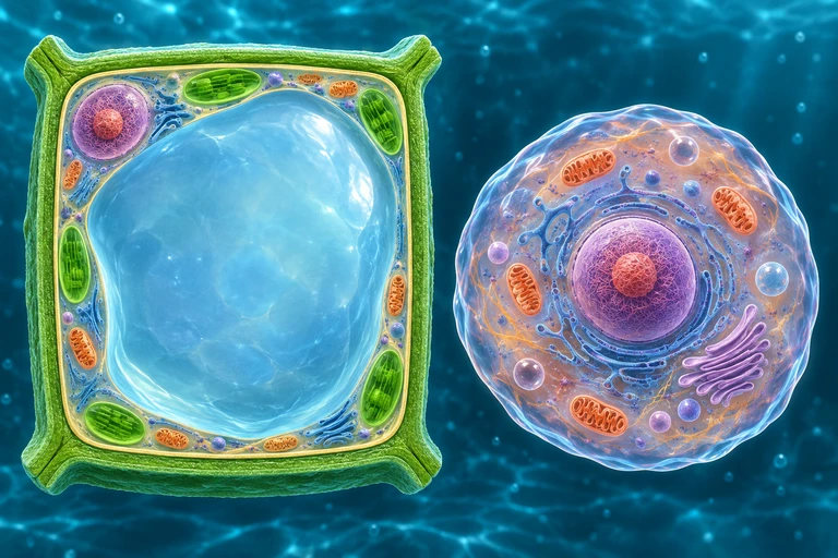 For nonpenetrating solutes, a plant wall limits expansion as water enters. Water loss retracts the protoplast from the wall during plasmolysis; animal cells lack this rigid wall.
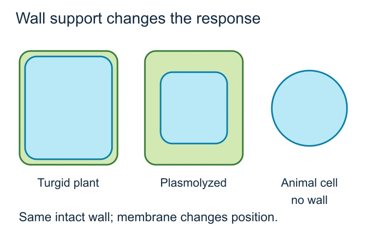 For nonpenetrating solutes, a plant wall limits expansion as water enters. Water loss retracts the protoplast from the wall during plasmolysis; animal cells lack this rigid wall.
SECTION 5
Organelle Classification and Comparison
One of the most frequently tested topics on the AP Biology exam is the comparison between prokaryotic and eukaryotic cells, as well as the distinguishing features of plant versus animal cells. The table below provides a systematic comparison of key structures and their presence or absence across these three categories.
| Structure / Feature | Prokaryotic Cell | Animal Cell | Plant Cell |
|---|---|---|---|
| Plasma membrane | Present | Present | Present |
| Cell wall | Present (peptidoglycan) | Absent | Present (cellulose) |
| Nucleus (membrane-bound) | Absent (nucleoid region) | Present | Present |
| Mitochondria | Absent | Present | Present |
| Chloroplasts | Absent | Absent | Present |
| Central vacuole | Absent | Absent (small vacuoles) | Present (large) |
| Ribosomes | 70S | 80S (70S in organelles) | 80S (70S in organelles) |
| DNA structure | Circular, no histones | Linear, with histones | Linear, with histones |
| Cytoskeleton | Limited (FtsZ, MreB) | Extensive | Extensive |
Comparison of prokaryotic, animal, and plant cell features
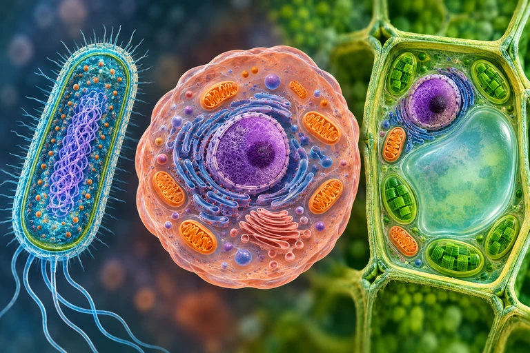 Bacteria, animal cells and photosynthetic leaf cells share a plasma membrane, DNA and ribosomes. The bacterium lacks a membrane-bound nucleus; both eukaryotic examples contain mitochondria, and the leaf cell also contains chloroplasts.
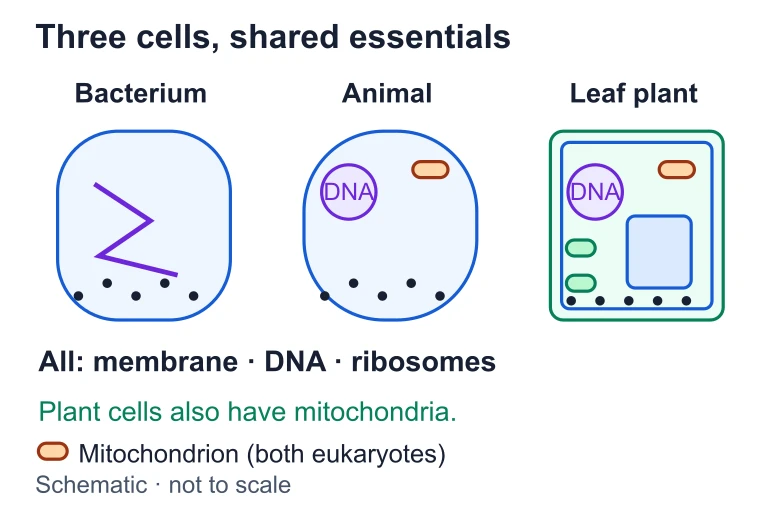 Bacteria, animal cells and photosynthetic leaf cells share a plasma membrane, DNA and ribosomes. The bacterium lacks a membrane-bound nucleus; both eukaryotic examples contain mitochondria, and the leaf cell also contains chloroplasts.
SECTION 6
Worked Example — Water Potential Calculation
Water potential calculations are a staple of the AP Biology exam, appearing frequently in both multiple-choice and free-response questions. The following worked example walks through a typical problem involving osmosis in plant cells.
Worked example
- 1
Step 1 — Identify the Given Information
A plant cell is placed in a 0.3 M sucrose solution at 25 °C. The cell's Ψₚ (pressure potential) is 0.5 bar. Sucrose is a non-ionizing solute (i = 1). We need to determine the cell's total water potential and predict the direction of water movement. - 2
Step 2 — Convert Temperature to Kelvin
T = 25 °C + 273 = 298 KT = 298 K - 3
Step 3 — Calculate Solute Potential (Ψₛ)
Using Ψₛ = −iCRT: Ψₛ = −(1)(0.3 mol/L)(0.0831 L·bar/mol·K)(298 K) = −(1)(0.3)(24.76) = −7.43 barΨₛ = −7.43 bar - 4
Step 4 — Calculate Total Water Potential (Ψ)
Ψ = Ψₛ + Ψₚ = −7.43 bar + 0.5 bar = −6.93 barΨ = −6.93 bar - 5
Step 5 — Determine the Water Potential of the Surrounding Solution
For an open container of 0.3 M sucrose, Ψₚ = 0 (no physical pressure is applied), so Ψ = Ψₛ + 0 = −7.43 bar. The surrounding solution's Ψ = −7.43 bar.Ψ(solution) = −7.43 bar - 6
Step 6 — Predict the Direction of Water Movement
Water moves from high Ψ to low Ψ. The cell's Ψ (−6.93 bar) is higher than the solution's Ψ (−7.43 bar), so water will move out of the cell and into the surrounding solution. The cell will lose turgor and begin to plasmolyze.Water flows OUT of the cell → plasmolysis
SECTION 7
The Endomembrane System — Functions and Interconnections
The endomembrane system is one of the defining features of eukaryotic cells, and it is a concept that the AP exam tests extensively. This system consists of the nuclear envelope, endoplasmic reticulum (both rough and smooth), Golgi apparatus, lysosomes, vacuoles, and the plasma membrane — organelles that either share membranes or exchange membrane segments via vesicular transport. Understanding how these components interact to synthesize, modify, package, and secrete proteins is essential for multiple AP exam topics, including signal transduction and cellular communication.
| Component | Primary Functions | Key Details for AP Exam |
|---|---|---|
| Nuclear Envelope | Separates chromatin from cytoplasm; regulates molecular transport via nuclear pores | Double membrane continuous with rough ER; nuclear pore complexes regulate import/export of mRNA, ribosomal subunits, and transcription factors |
| Rough ER | Synthesis of secretory proteins, membrane proteins, and glycoproteins; initial folding and quality control | Studded with ribosomes; signal peptide on nascent polypeptide directs ribosome to ER membrane; proteins enter ER lumen cotranslationally |
| Smooth ER | Lipid synthesis, detoxification of drugs and poisons, calcium ion storage | Abundant in liver cells (detoxification) and muscle cells (Ca²⁺ storage in sarcoplasmic reticulum); lacks ribosomes |
| Golgi Apparatus | Modification (glycosylation, phosphorylation), sorting, and packaging of proteins into vesicles | Has cis (receiving) and trans (shipping) faces; receives transport vesicles from ER at cis face; dispatches vesicles from trans face to plasma membrane, lysosomes, or back to ER |
| Lysosomes | Intracellular digestion of macromolecules, damaged organelles (autophagy), and engulfed particles (phagocytosis) | Contain hydrolytic enzymes active at pH ≈ 5; lysosomal membrane contains H⁺ pumps; Tay-Sachs disease results from a defective lysosomal enzyme |
Components of the endomembrane system and their relevance to the AP Biology exam
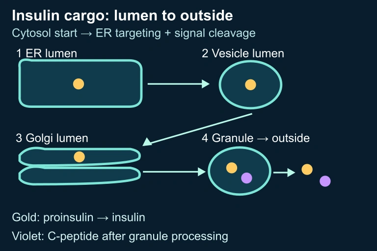 Lumen route and processing schematic
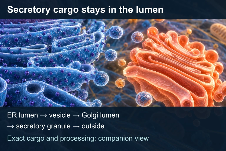 Translation begins in the cytosol and is targeted to the ER, where the signal peptide is cleaved. Secretory cargo stays in noncytosolic lumens as it travels through the Golgi and granules. Granule processing produces insulin and C-peptide, which are released outside.
SECTION 8
Connections to Advanced Topics
Cell structure and function connects directly to nearly every other unit on the AP Biology exam. A deep understanding of organelle function is prerequisite for cellular energetics (Units 3 and 4), where the mitochondrial matrix and inner membrane are the sites of the citric acid cycle and oxidative phosphorylation, respectively, and the chloroplast thylakoids and stroma are the sites of light reactions and the Calvin cycle. Similarly, the nucleus and ribosome are central to gene expression (Unit 6), while the plasma membrane and its receptors are central to cell communication (Unit 4). The table below maps key connections between cell structure and advanced topics tested on the AP exam.
| Cell Structure Concept | Advanced AP Biology Connection |
|---|---|
| Mitochondrial structure | Chemiosmosis and the electron transport chain require the inner mitochondrial membrane's impermeability to H⁺ and the intermembrane space for the proton gradient (ΔpH) |
| Chloroplast structure | Thylakoid membrane organization enables light-dependent reactions; stroma is the site of carbon fixation (Calvin cycle) |
| Plasma membrane receptors | G protein-coupled receptors and receptor tyrosine kinases initiate signal transduction cascades (Unit 4); ligand binding triggers conformational changes |
| Cytoskeleton | Microtubule spindle fibers are essential for chromosome segregation during mitosis and meiosis (Units 5 and 6); actin microfilaments drive cytokinesis and cell motility |
| Endosymbiotic origin | Evidence for evolution (Unit 7): homologous structures (double membranes, own DNA), phylogenetic analysis of rRNA genes supports common ancestry with alpha-proteobacteria (mitochondria) and cyanobacteria (chloroplasts) |
How cell structure concepts connect to other AP Biology units
As you progress through the AP Biology curriculum, continually revisit cell structure concepts. The same organellar architecture you learn here will reappear in the context of energy transformations, gene regulation, heredity, and evolution. Recognizing these connections will improve not only your free-response answers but also your ability to reason through unfamiliar experimental scenarios — a skill the redesigned AP exam increasingly rewards.
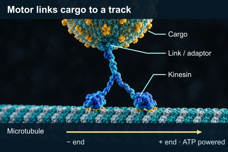 A kinesin motor and adaptor connect a cargo vesicle to a microtubule. ATP powers movement toward the plus end in this selected transport model.
SECTION 9
Practice Problems
Question 1: CONCEPTUAL
- A. Mitochondria are found in both plant and animal cells.
- B. Mitochondria contain their own circular DNA and replicate by binary fission.
- C. Mitochondria produce ATP through oxidative phosphorylation.
- D. Mitochondria are enclosed by a single phospholipid membrane.
Show answer (B)
Question 2: BASIC CALCULATION
- A. −12.27 bar
- B. −24.53 bar
- C. −49.06 bar
- D. −6.14 bar
Show answer (B)
Question 3: INTERMEDIATE
- A. DNA replication in the nucleus would be inhibited.
- B. Secretory proteins would accumulate in the ER and fail to be properly modified and exported.
- C. ATP production by mitochondria would decrease significantly.
- D. The cell would immediately undergo apoptosis due to lysosomal rupture.
Show answer (B)
Question 4: APPLIED
8 ptsShow answer
Question 5: CRITICAL THINKING
4 pts