
SECTION 1
Historical Context & Motivation
The realization that cells are not homogeneous bags of enzymes, but rather intricately organized structures with distinct internal compartments, ranks among the most transformative insights in modern biology. Early microscopists could distinguish a nucleus from the surrounding cytoplasm, yet the true extent of cell compartmentalization only became apparent with the advent of electron microscopy in the mid-twentieth century. The discovery that eukaryotic cells harbor dozens of membrane-bound organelles—each maintaining its own pH, redox state, and enzymatic repertoire—raised a fundamental question: why would evolution favor such elaborate internal architecture over the simpler, open cytoplasmic plan of prokaryotes?
1665 • Hooke's "Cells"
1831 • Discovery of the Nucleus
1898 • Golgi Apparatus Described
1945 • Cell Fractionation
1967 • Endosymbiotic Theory
These milestones converge on a central question that drives this lesson: How does the partitioning of eukaryotic cells into membrane-bound compartments enable metabolic efficiency, regulatory precision, and the evolution of multicellular complexity? Understanding compartmentalization is essential for the AP Biology exam, where it underpins concepts ranging from cellular respiration and photosynthesis to signal transduction and gene expression.
SECTION 2
Core Principles of Compartmentalization
Compartmentalization refers to the organization of cellular contents into distinct, membrane-enclosed regions that maintain unique internal environments. This principle is fundamentally tied to the properties of biological membranes—selectively permeable phospholipid bilayers embedded with transport proteins, receptors, and enzymes. Because membranes restrict the free diffusion of ions and polar molecules, each organelle can establish and maintain conditions that differ dramatically from those in the cytosol. Several foundational ideas underpin this concept.
Selective Permeability
Increased Surface Area
Concentration of Reactants
Separation of Incompatible Processes
Regulation via Gated Transport
KEY TAKEAWAY
SECTION 3
Visual Explanation — The Eukaryotic Cell Plan
The diagram above captures the essential spatial logic of eukaryotic compartmentalization. Notice that each organelle is bounded by at least one lipid bilayer, creating a topologically distinct lumen or matrix. The endomembrane system—comprising the endoplasmic reticulum (ER), Golgi apparatus, lysosomes, and transport vesicles—forms a functionally connected network in which proteins and lipids flow from one compartment to the next via budding and fusion of vesicles. In contrast, mitochondria and peroxisomes are not part of the endomembrane system; they acquire their proteins through direct import from the cytosol using signal sequences recognized by specific translocases. This distinction is critical for AP Biology: the endomembrane system relies on vesicular transport, whereas mitochondria and chloroplasts use translocon-mediated import.
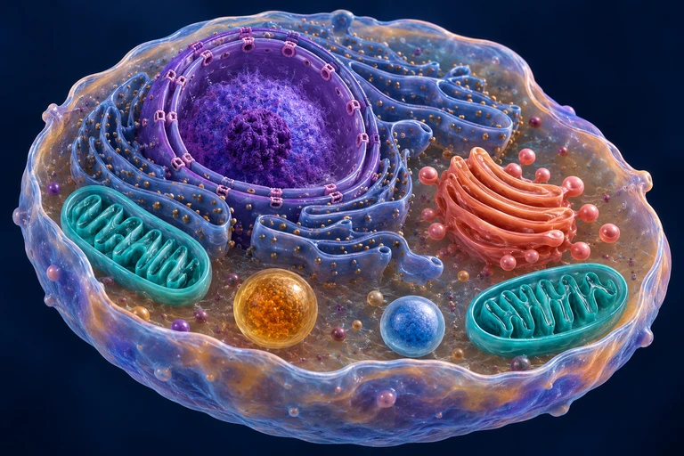 Membranes separate cytosol from organelle lumens and maintain distinct chemical environments. The nuclear outer membrane connects with the endoplasmic reticulum; mitochondrial and peroxisomal protein import follows routes distinct from ER–Golgi vesicle traffic.
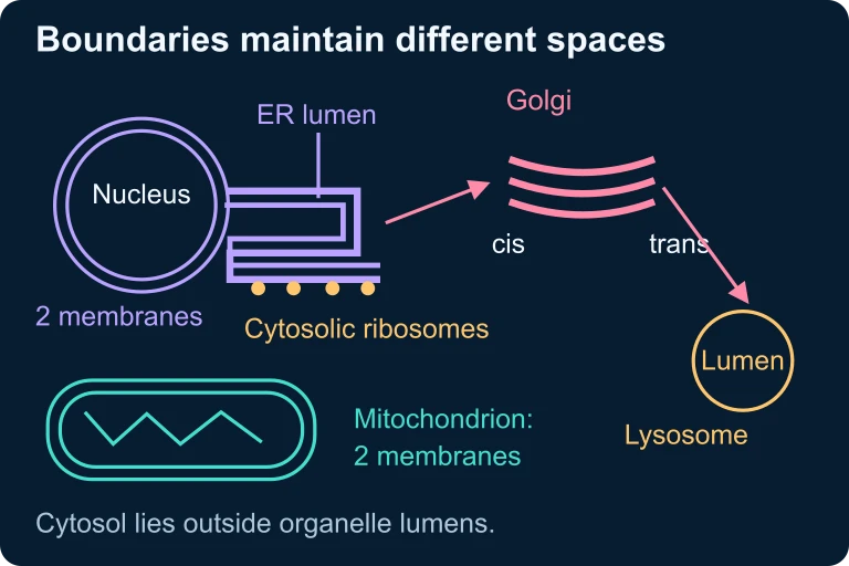 Membranes separate cytosol from organelle lumens and maintain distinct chemical environments. The nuclear outer membrane connects with the endoplasmic reticulum; mitochondrial and peroxisomal protein import follows routes distinct from ER–Golgi vesicle traffic.
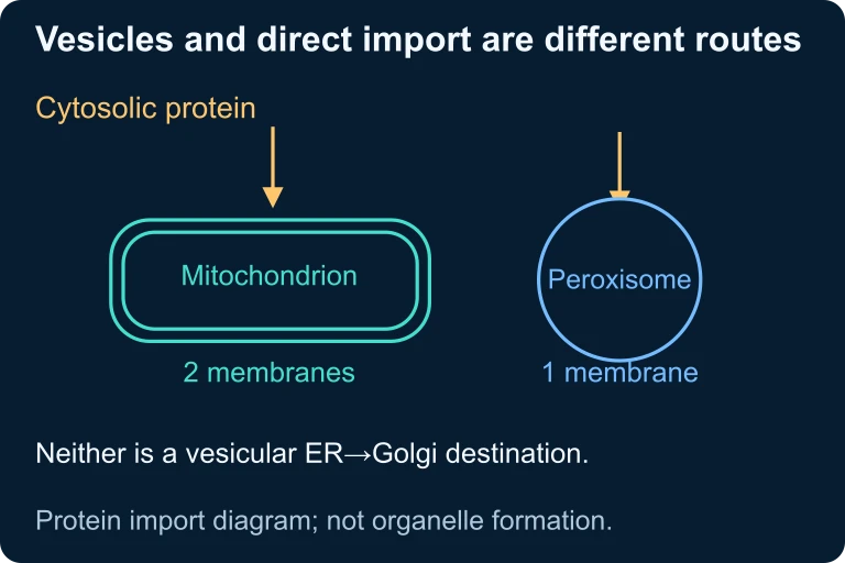 Mitochondrial and peroxisomal proteins can enter from the cytosol through direct-import routes distinct from ER–Golgi vesicle transport.
SECTION 4
Mechanisms of Compartmentalization
Because most organellar proteins are encoded by nuclear genes, the cell faces a fundamental logistics problem: how does a newly synthesized polypeptide reach its correct destination? Two major mechanisms solve this challenge, and both depend on signal sequences—short stretches of amino acids that function as molecular zip codes, directing each protein to its intended compartment.
Co-translational Targeting to the ER
Proteins destined for the endomembrane system, the plasma membrane, or secretion carry an N-terminal ER signal peptide, typically 16–30 amino acids rich in hydrophobic residues. As the signal peptide emerges from the ribosome, it is recognized by the signal recognition particle (SRP), a ribonucleoprotein complex that pauses translation and docks the ribosome–nascent chain complex onto the SRP receptor at the ER membrane. Translation then resumes through the Sec61 translocon, a gated protein channel, threading the polypeptide directly into the ER lumen. Signal peptidase cleaves the signal peptide, and the protein undergoes folding, glycosylation, and quality control within the ER before being packaged into COPII-coated vesicles for transport to the Golgi.
Post-translational Import into Mitochondria
Mitochondrial matrix proteins are synthesized on free ribosomes in the cytosol and carry an N-terminal mitochondrial targeting sequence (MTS)—a positively charged, amphipathic helix. Cytosolic chaperones (primarily Hsp70) maintain the polypeptide in an unfolded, import-competent state. The MTS is recognized by receptors on the TOM complex (translocase of the outer membrane), passed through the TOM pore, and then transferred to the TIM23 complex (translocase of the inner membrane). The electrochemical gradient (Δψ) across the inner membrane and matrix Hsp70 pulling activity drive translocation into the matrix, where the MTS is cleaved by mitochondrial processing peptidase.
Maintaining Compartment Identity
Beyond protein targeting, compartments actively maintain their unique environments. Lysosomes sustain a luminal pH of approximately 4.5–5.0 through the action of V-type H⁺-ATPases that pump protons against their concentration gradient, consuming ATP in the process. The ER maintains an oxidizing environment to promote disulfide bond formation, while the cytosol remains reducing—conditions that would be mutually destructive if not separated by the ER membrane. Peroxisomes sequester hydrogen peroxide–generating oxidases away from the cytosol, containing the reactive oxygen species within their catalase-rich lumen. Each of these examples illustrates the same overarching principle: membrane boundaries enable each compartment to establish and defend a specialized chemical milieu.
AP EXAM TIP
 A lysosomal lumen at pH 4.8 has about 251 times the hydrogen-ion concentration of cytosol at pH 7.2. A V-type H⁺-ATPase uses ATP to pump protons into the lumen and maintain this gradient.
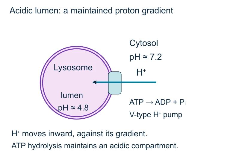 A lysosomal lumen at pH 4.8 has about 251 times the hydrogen-ion concentration of cytosol at pH 7.2. A V-type H⁺-ATPase uses ATP to pump protons into the lumen and maintain this gradient.
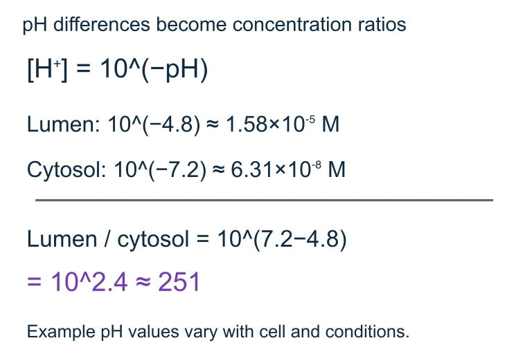 Hydrogen-ion concentration varies as 10 to the negative pH. The lumen-to-cytosol ratio is 10^(7.2−4.8), or about 251.
SECTION 5
Organelle-by-Organelle Breakdown
To appreciate how compartmentalization supports cellular function, it is useful to examine each major organelle as a specialized microenvironment. The table below summarizes the key features—number of bounding membranes, internal pH, primary functions, and distinguishing characteristics—that the AP exam expects you to know.
| Organelle | Membranes | Luminal pH | Primary Functions |
|---|---|---|---|
| Nucleus | Double (nuclear envelope) | ≈ 7.2 | DNA replication, transcription, ribosome assembly (nucleolus) |
| Rough ER | Single (continuous with nuclear envelope) | ≈ 7.2 | Co-translational protein import, folding, N-linked glycosylation |
| Smooth ER | Single | ≈ 7.2 | Lipid synthesis, Ca²⁺ storage, drug detoxification |
| Golgi Apparatus | Single (stacked cisternae) | cis ≈ 6.7 → trans ≈ 6.0 | Glycoprotein modification, sorting, vesicle packaging |
| Lysosome | Single | ≈ 4.5–5.0 | Intracellular digestion via acid hydrolases, autophagy |
| Mitochondrion | Double (outer + inner with cristae) | Matrix ≈ 7.8; IMS ≈ 7.0 | Krebs cycle (matrix), oxidative phosphorylation (inner membrane), apoptosis regulation |
| Chloroplast | Double + thylakoid membrane | Stroma ≈ 8.0; thylakoid lumen ≈ 5.0 | Light reactions (thylakoid membrane), Calvin cycle (stroma) |
| Peroxisome | Single | ≈ 7.0 | Fatty acid β-oxidation, H₂O₂ detoxification by catalase |
Summary of major eukaryotic organelles and their compartment-specific properties.
The pH spectrum diagram powerfully illustrates a critical point: compartmentalization is not merely about spatial separation but about energetic investment. Maintaining a lysosomal pH of 4.5 in a cytosol at pH 7.2 requires V-type ATPases to continuously pump protons against a steep electrochemical gradient. Similarly, the proton gradient across the inner mitochondrial membrane, generated by the electron transport chain, is the very energy source that drives ATP synthase. In both cases, the membrane boundary is essential: without it, protons would equilibrate and the functional gradient would collapse. This perspective connects compartmentalization directly to bioenergetics, a major theme of the AP Biology curriculum.
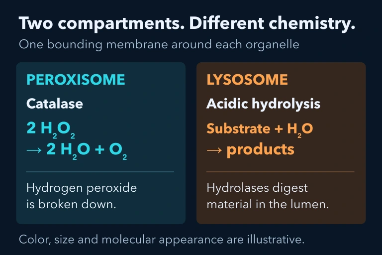 Peroxisomal catalase converts hydrogen peroxide to water and oxygen. Lysosomal acidic hydrolases perform a different digestive role in a separate single-membrane compartment.
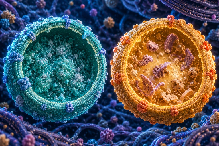 Peroxisomal catalase converts hydrogen peroxide to water and oxygen. Lysosomal acidic hydrolases perform a different digestive role in a separate single-membrane compartment.
SECTION 6
Worked Example — Tracing a Secreted Protein
One of the most common AP Biology questions asks you to trace the path of a secreted protein—such as insulin—from gene to extracellular release. This worked example walks through each compartment the protein visits, emphasizing the role of compartmentalization at every step.
Tracing Insulin from Gene to Secretion
- 1
Step 1 — Transcription in the Nucleus
The insulin gene (INS) is transcribed by RNA polymerase II within the nucleus. The pre-mRNA is processed (5' capping, splicing, polyadenylation) and exported through nuclear pore complexes to the cytoplasm.Compartment: Nucleus → Cytoplasm - 2
Step 2 — Translation & Co-translational Import into the Rough ER
Free ribosomes begin translating the mRNA. The emerging N-terminal signal peptide is recognized by SRP, which directs the ribosome to the rough ER membrane. Translation continues through the Sec61 translocon, threading preproinsulin into the ER lumen. Signal peptidase cleaves the signal peptide, yielding proinsulin. Disulfide bonds form in the oxidizing ER environment, and the protein undergoes quality control.Compartment: Cytoplasm → Rough ER lumen - 3
Step 3 — COPII Vesicle Transport to the Golgi
Properly folded proinsulin is packaged into COPII-coated vesicles that bud from ER exit sites. These vesicles fuse with the cis face of the Golgi apparatus. Properly folded proinsulin passes through the Golgi and is sorted at the trans-Golgi network into immature secretory granules. Normal insulin biosynthesis does not require glycosylating proinsulin.Compartment: ER → Golgi (cis → trans) - 4
Step 4 — Proteolytic Processing in Secretory Granules
At the trans-Golgi network, proinsulin is sorted into regulated secretory granules. Within these acidic vesicles, prohormone convertases cleave proinsulin to produce mature insulin (A and B chains connected by disulfide bonds) plus the C-peptide. The granules are stored in the cytoplasm, awaiting the appropriate signal.Compartment: trans-Golgi → Secretory granule - 5
Step 5 — Exocytosis
When blood glucose rises, a signaling cascade triggers the secretory granules to move along the cytoskeleton to the plasma membrane. SNARE proteins mediate membrane fusion, releasing mature insulin into the extracellular space via regulated exocytosis. Throughout this entire journey, insulin was never free in the cytosol—compartmentalization ensured it moved through a continuous membrane-enclosed pathway.Compartment: Secretory granule → Extracellular space
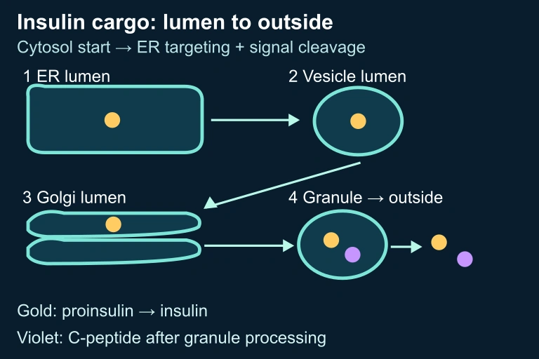 Lumen route and processing schematic
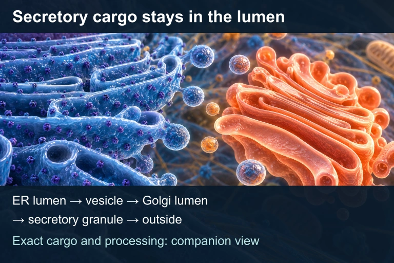 Translation begins in the cytosol and is targeted to the ER, where the signal peptide is cleaved. Secretory cargo stays in noncytosolic lumens as it travels through the Golgi and granules. Granule processing produces insulin and C-peptide, which are released outside.
KEY TAKEAWAY
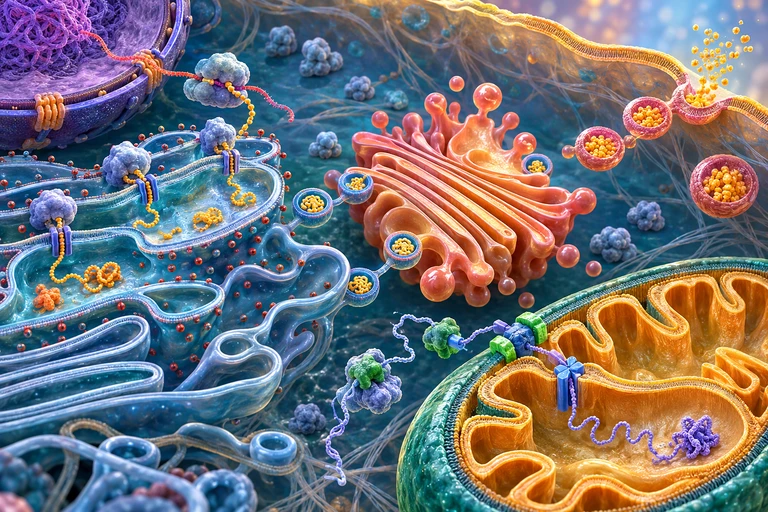 Secretory cargo stays inside membrane-enclosed spaces as vesicles bud and fuse. Proteins entering the mitochondrial matrix follow a separate direct-import route.
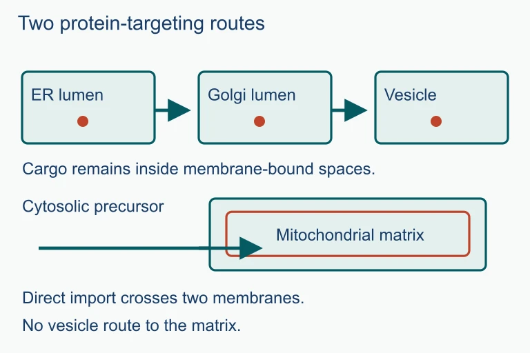 Secretory cargo stays inside membrane-enclosed spaces as vesicles bud and fuse. Proteins entering the mitochondrial matrix follow a separate direct-import route.
SECTION 7
Prokaryotic vs. Eukaryotic Organization
Compartmentalization is the defining structural difference between prokaryotic and eukaryotic cells, and AP Biology frequently tests your ability to compare these two organizational strategies. Prokaryotes are not devoid of internal organization—they employ protein-based microcompartments, lipid rafts, and localized enzyme complexes—but they lack the membrane-bound organelles that characterize eukaryotes. This distinction has profound consequences for genome size, metabolic versatility, and the evolution of multicellularity.
| Feature | Prokaryotic Cell | Eukaryotic Cell |
|---|---|---|
| Membrane-bound nucleus | Absent; DNA in nucleoid region | Present; DNA enclosed by nuclear envelope |
| Endomembrane system | Absent | ER, Golgi, lysosomes, vesicles |
| Electron transport | Plasma membrane | Inner mitochondrial membrane / thylakoid membrane |
| Transcription & translation | Coupled (co-transcriptional translation) | Separated by nuclear envelope; transcription in nucleus, translation in cytoplasm |
| Gene regulation | Primarily transcriptional (operons) | Multi-level: chromatin remodeling, transcription, RNA processing, translation, post-translational |
| Typical cell diameter | 0.5–5 μm | 10–100 μm |
Key structural and functional differences arising from compartmentalization.
WHY THIS MATTERS
SECTION 8
Evolutionary Origins — Endosymbiosis and Beyond
How did eukaryotic compartmentalization evolve? Two major hypotheses address different aspects of this question. The endosymbiotic theory, championed by Lynn Margulis and now supported by overwhelming molecular evidence, explains the origin of mitochondria and chloroplasts as formerly free-living α-proteobacteria and cyanobacteria, respectively, that were engulfed by an ancestral eukaryotic host cell and retained as endosymbionts. The autogenous hypothesis proposes that the nuclear envelope and endomembrane system arose from infoldings of the ancestral plasma membrane, progressively internalizing membrane-associated processes.
| Evidence | Endosymbiotic Theory (Mitochondria / Chloroplasts) | Autogenous Hypothesis (Endomembrane System) |
|---|---|---|
| Own DNA | Circular genome resembling bacterial DNA; encodes rRNAs, tRNAs, and some proteins | ER/Golgi/lysosomes lack their own genomes |
| Double membrane | Inner membrane derived from engulfed bacterium; outer from host's phagocytic vesicle | Nuclear envelope is a double membrane continuous with ER, consistent with plasma membrane invagination |
| Ribosome size | 70S ribosomes (bacterial-type), sensitive to chloramphenicol | Not applicable |
| Binary fission | Mitochondria and chloroplasts divide by fission, independent of cell cycle | Endomembrane compartments grow by vesicle budding and fusion |
| Phylogenetic support | Mitochondrial rRNA sequences cluster with α-proteobacteria; chloroplast sequences with cyanobacteria | Some archaeal membrane-bending proteins (ESCRT-III) found in eukaryotes, suggesting shared ancestry for membrane remodeling |
Key lines of evidence supporting two complementary models for the origin of eukaryotic compartmentalization.
These two hypotheses are not mutually exclusive—they address the origins of different compartments. What they share is the implication that compartmentalization was a prerequisite for the dramatic expansion of eukaryotic genome size, cell size, and functional complexity. Without the energy efficiency provided by endosymbiotic mitochondria and the regulatory sophistication enabled by the nuclear envelope, the transition to multicellularity may never have occurred. For the AP exam, be prepared to cite specific lines of evidence for endosymbiosis (double membrane, circular DNA, 70S ribosomes, binary fission) and to explain how the nuclear envelope enabled new forms of gene regulation.
SECTION 9
Practice Problems
Question 1: CONCEPTUAL
- A. It prevents ribosomes from degrading mRNA before transcription is complete.
- B. It allows RNA processing events such as splicing to occur before translation, enabling the production of multiple protein variants from a single gene.
- C. It increases the rate of translation by concentrating ribosomes near the DNA template.
- D. It ensures that all mRNA molecules are translated immediately after they are produced.
Show answer (B)
Question 2: BASIC CALCULATION
- A. 2.4-fold
- B. 24-fold
- C. 250-fold
- D. 2,400-fold
Show answer (C)
Question 3: INTERMEDIATE
- A. Insulin mRNA would fail to be transcribed from the INS gene.
- B. Preproinsulin would be translated on free ribosomes and remain in the cytosol rather than entering the ER lumen.
- C. Insulin would accumulate in the mitochondrial matrix due to mistargeting.
- D. The Golgi apparatus would be unable to glycosylate any proteins.
Show answer (B)
Question 4: APPLIED
4 ptsShow answer
Question 5: CRITICAL THINKING
4 ptsShow answer
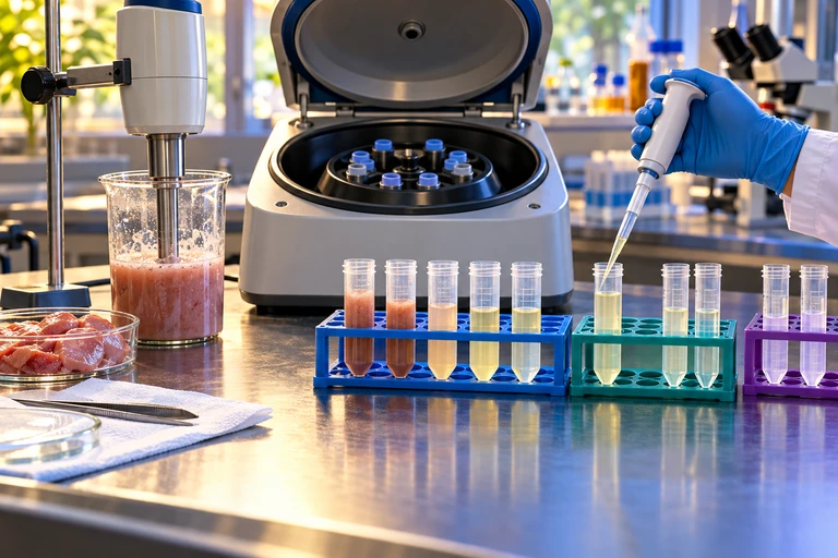 Each centrifugation step processes the previous supernatant while retaining its pellet. Marker activities identify enriched fractions rather than pure organelles; mitochondrial and lysosomal markers both peak in fraction 2.
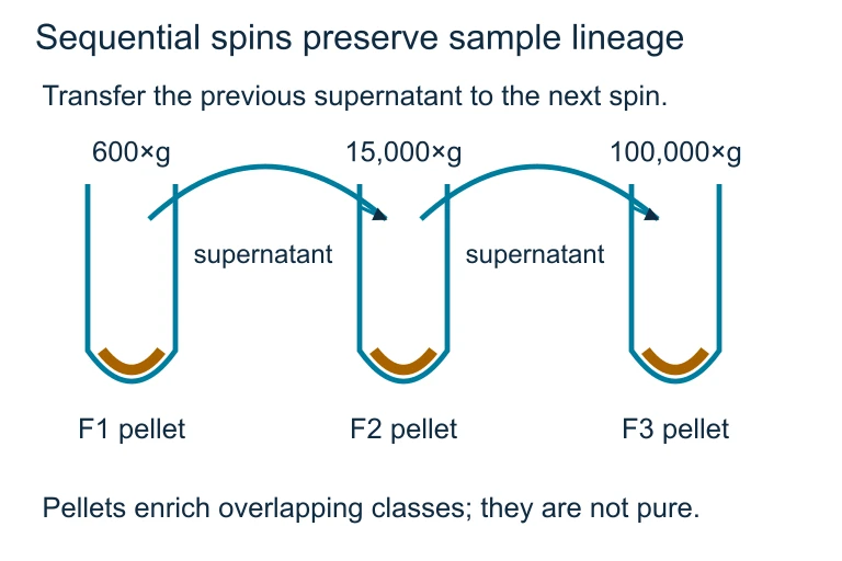 Each centrifugation step processes the previous supernatant while retaining its pellet. Marker activities identify enriched fractions rather than pure organelles; mitochondrial and lysosomal markers both peak in fraction 2.
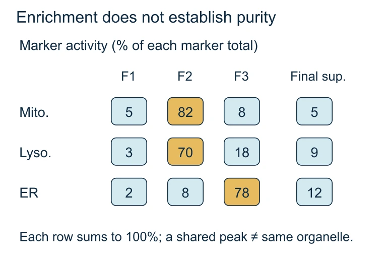 Each marker-activity row sums to 100%. Shared mitochondrial and lysosomal peaks in fraction 2 show that enrichment does not establish purity.
SUMMARY
Cell Compartmentalization — Key Concepts Review
Summary
Cell compartmentalization is the organization of eukaryotic cells into distinct membrane-bound organelles, each maintaining a specialized chemical environment. The selective permeability of phospholipid bilayers enables compartments to establish unique pH, redox conditions, and enzymatic compositions. Key mechanisms include signal sequences that direct proteins to the correct organelle, vesicular transport through the endomembrane system (ER → Golgi → lysosome / plasma membrane), and translocon-mediated import into mitochondria and chloroplasts.
Compartmentalization confers major advantages: it increases metabolic efficiency by concentrating reactants, separates incompatible reactions (e.g., lysosomal hydrolysis from cytosolic biosynthesis), and enables multi-level gene regulation by separating transcription from translation via the nuclear envelope. Evolutionarily, the endosymbiotic origin of mitochondria and chloroplasts, supported by their double membranes, circular DNA, 70S ribosomes, and phylogenetic evidence, explains how some of the most critical compartments arose. Mastery of compartmentalization connects to nearly every major AP Biology topic—from cellular energetics and protein trafficking to cell signaling and disease.