
SECTION 1
Historical Context & Motivation
Long before chemists understood the molecular basis of nutrition, humans recognized that starchy and sweet foods provided rapid energy. The scientific study of carbohydrates began in earnest during the early nineteenth century, when chemists noticed that many plant-derived compounds shared a curious empirical formula—carbon atoms appeared to be "hydrated" in a fixed ratio with water molecules, giving rise to the name carbo-hydrate. Although we now know the name is chemically misleading (these molecules are not literally hydrated carbon), it persists as a testament to the field's historical roots. Understanding how scientists progressively dissected sugars from simple glucose to massive polysaccharides reveals how structure–function relationships became a central theme in modern biology.
1811 • Kirchhoff Converts Starch to Sugar
1838 • Anselme Payen Isolates Cellulose
1891 • Emil Fischer Elucidates Sugar Stereochemistry
1929 • Haworth Determines Ring Structures
1953 • Glycobiology Emerges
This historical trajectory highlights a central question that the AP Biology curriculum revisits: how does the arrangement of atoms within carbohydrate monomers dictate the emergent properties of the polymers they form? The answer lies in the interplay between stereochemistry, glycosidic linkage type, and branching patterns—concepts we will explore in the sections that follow.
SECTION 2
Core Principles & Definitions
Carbohydrates are organic molecules composed of carbon, hydrogen, and oxygen, typically in a ratio approximating Cn(H2O)n. They serve as primary energy sources, structural materials, and molecular identification tags. The biological diversity of carbohydrates emerges from a few foundational principles that govern their synthesis, classification, and function.
Monosaccharides
Glycosidic Linkages
Disaccharides & Polysaccharides
Dehydration Synthesis & Hydrolysis
Structure Determines Function
SECTION 3
Visual Explanation — Monosaccharide Structure
The diagram above illustrates a critical concept for AP Biology: in solution, monosaccharides rarely exist as straight chains. Instead, an intramolecular reaction between the carbonyl carbon (C1 in aldoses) and a downstream hydroxyl group closes the molecule into a stable ring. For glucose, this produces a six-membered pyranose ring containing five carbons and one oxygen. The orientation of the hydroxyl group on C1 distinguishes the α anomer (OH below the ring plane) from the β anomer (OH above the ring plane). This seemingly minor stereochemical difference—α versus β—has profound biological consequences: it determines whether enzymatic machinery can digest the polymer (starch, with α-linkages, is digestible by humans; cellulose, with β-linkages, is not).
SECTION 4
Dehydration Synthesis & Hydrolysis
All carbohydrate polymers are assembled by a single class of reaction—dehydration synthesis (also called condensation)—and disassembled by hydrolysis. Understanding the thermodynamic and mechanistic logic of these reactions is essential because the same logic governs peptide bond formation in proteins, phosphodiester bond formation in nucleic acids, and ester bond formation in lipids. The universality of dehydration/hydrolysis is one of biology's most elegant unifying themes.
DEHYDRATION SYNTHESIS (GLYCOSIDIC BOND FORMATION)
A hydroxyl group (−OH) from one sugar and a hydrogen (H−) from the hydroxyl of another are removed, releasing one water molecule. The resulting glycosidic bond (−O−) links the two residues. This reaction is endergonic and coupled to ATP hydrolysis in vivo.
HYDROLYSIS (GLYCOSIDIC BOND CLEAVAGE)
Water is consumed to break the glycosidic bond, regenerating two free monosaccharides (or smaller oligosaccharides). This reaction is catalyzed by specific enzymes: amylase hydrolyzes starch, lactase hydrolyzes lactose, and cellulase (produced by certain microbes) hydrolyzes cellulose.
The specificity of the glycosidic bond is described by the carbons involved and the anomeric configuration. For instance, maltose contains an α-1,4-glycosidic linkage (α orientation, connecting C1 of one glucose to C4 of the next), while cellobiose contains a β-1,4-glycosidic linkage. The α versus β distinction is the critical variable that enzymes recognize. Human digestive enzymes possess active sites complementary to α-glycosidic bonds but cannot accommodate the geometry of β-glycosidic bonds—which is precisely why we can digest starch but not cellulose, even though both are glucose polymers.
SECTION 5
Polysaccharide Classification & Comparison
Polysaccharides are the most structurally and functionally diverse carbohydrates, and the AP Biology exam expects you to distinguish among the major types based on monomer identity, linkage type, branching pattern, and biological role. The following diagram and table provide a comprehensive classification of the four polysaccharides you must know.
| Polysaccharide | Monomer | Linkage | Branching | Function | Found In |
|---|---|---|---|---|---|
| Starch | α-glucose | α-1,4 (amylose); α-1,4 + α-1,6 (amylopectin) | None (amylose); moderate (amylopectin) | Energy storage | Plants (tubers, seeds) |
| Glycogen | α-glucose | α-1,4 + α-1,6 | Extensive (every 8–12 residues) | Energy storage | Animals (liver, muscle) |
| Cellulose | β-glucose | β-1,4 | None (strictly linear) | Structural support | Plant cell walls |
| Chitin | N-acetylglucosamine (NAG) | β-1,4 | None (strictly linear) | Structural support | Arthropod exoskeletons; fungal cell walls |
Summary of the four polysaccharides emphasized on the AP Biology exam
Note a recurring pattern in the table: energy-storage polysaccharides use α-linkages and are often branched, providing many nonreducing ends from which enzymes can simultaneously release glucose monomers for rapid mobilization. In contrast, structural polysaccharides use β-linkages with no branching, allowing chains to align in parallel and form extensive hydrogen-bond networks that confer tensile strength. Glycogen is more heavily branched than amylopectin because animals require faster glucose mobilization during bursts of muscular activity, whereas plants can rely on slower, steady hydrolysis of starch granules.
 Amylose, amylopectin and glycogen use α-linked glucose chains; amylopectin and glycogen also have α-1,6 branches. Cellulose and chitin use unbranched β-1,4 backbones.
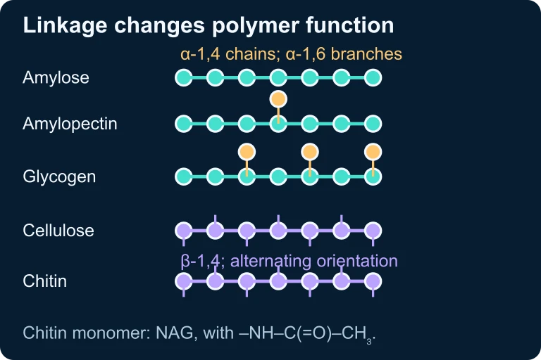 Amylose, amylopectin and glycogen use α-linked glucose chains; amylopectin and glycogen also have α-1,6 branches. Cellulose and chitin use unbranched β-1,4 backbones.
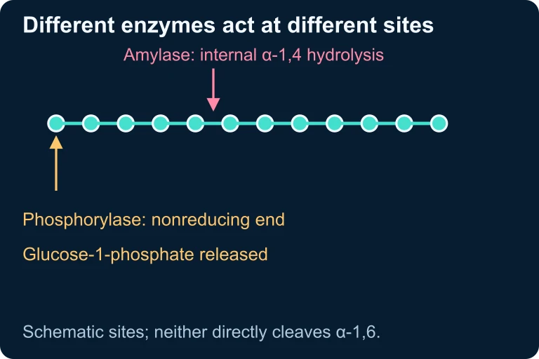 Amylase hydrolyzes internal α-1,4 bonds. Glycogen phosphorylase acts at nonreducing ends to release glucose-1-phosphate; neither enzyme directly cleaves α-1,6 branch bonds.
SECTION 6
Worked Example — Polymer Assembly & Water Release
A common AP Biology problem asks you to reason about the relationship between the number of monomers in a polymer and the number of water molecules released (or consumed) during synthesis (or hydrolysis). Let us work through a representative example.
Worked example
- 1
Step 1 — Identify the Reaction Type
Each glycosidic bond is formed via dehydration synthesis, which releases one molecule of H₂O per bond formed. - 2
Step 2 — Count the Bonds
A linear polymer of n monomers contains (n − 1) glycosidic bonds. For 500 glucose residues in a linear chain: 500 − 1 = 499 glycosidic bonds.499 bonds - 3
Step 3 — Calculate Water Released
Since each bond formation releases one H₂O: water molecules released = 499.499 molecules of H₂O released - 4
Step 4 — Consider Branching (Extension)
A connected, acyclic polysaccharide containing 500 glucose residues has 499 glycosidic bonds regardless of branching. If 50 of those bonds are α(1→6) branch linkages, the remaining 449 are α(1→4) linkages. In this lesson's net dehydration model, assembly releases 499 water molecules.499 H₂O; branching changes connectivity, not the total n − 1 bond count.
SECTION 7
Energy Storage vs. Structural Support — Functional Comparison
One of the most frequently tested concepts on the AP Biology exam is the ability to distinguish between carbohydrates optimized for energy storage and those optimized for structural support. The table below organizes the key functional trade-offs.
| Feature | Energy-Storage Carbohydrates (Starch, Glycogen) | Structural Carbohydrates (Cellulose, Chitin) |
|---|---|---|
| Linkage type | α-glycosidic | β-glycosidic |
| Chain geometry | Helical coils (compact) | Straight, parallel chains |
| Branching | Often branched (especially glycogen) | Unbranched |
| Interchain bonding | Minimal; chains are relatively independent | Extensive hydrogen bonding between parallel chains → microfibrils |
| Solubility | Somewhat soluble or forms colloidal suspensions | Insoluble in water |
| Enzymatic accessibility | Easily hydrolyzed by amylases and phosphorylases | Requires cellulase (absent in most animals) |
| Biological advantage | Rapid glucose mobilization; compact packing | High tensile strength; resistance to enzymatic degradation |
Functional comparison of storage vs. structural polysaccharides
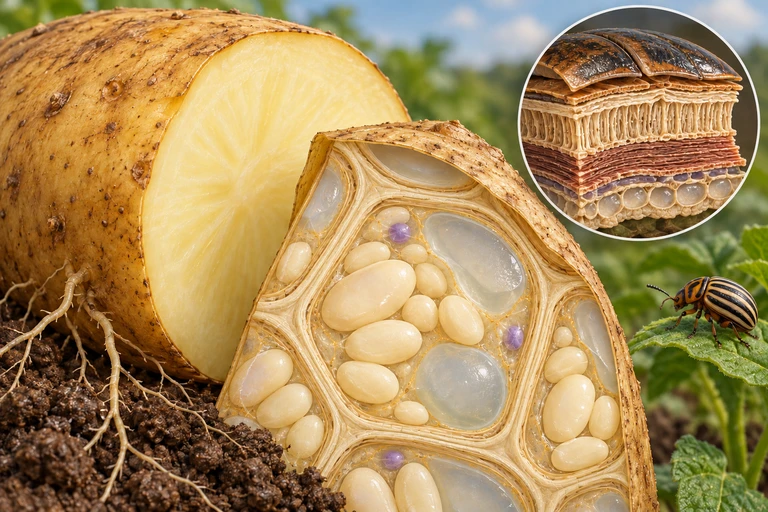
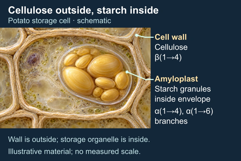 In potato storage cells, cellulose supports the extracellular wall while starch is stored inside amyloplasts. The enlarged structures are schematic.
SECTION 8
Connection to Advanced Topics — Glycobiology & Cell Signaling
While the AP Biology exam primarily tests your understanding of monosaccharides and the four major polysaccharides, it also touches on the role of carbohydrates in cell recognition and signaling. The surface of virtually every eukaryotic cell is coated with a dense layer of carbohydrate chains called the glycocalyx. These carbohydrates are covalently attached to membrane proteins (glycoproteins) and membrane lipids (glycolipids), and they face the extracellular environment where they serve as molecular identity tags.
| AP Biology Focus | Advanced Connection |
|---|---|
| Monosaccharides as cellular fuel (glucose → glycolysis → cellular respiration) | Metabolic regulation: hexokinase traps glucose inside cells by phosphorylation; insulin/glucagon signaling controls blood glucose homeostasis |
| Polysaccharides as energy storage and structural materials | Glycogen storage diseases (GSDs) illustrate how defects in single enzymes disrupt polysaccharide metabolism with severe clinical consequences |
| Cell-surface carbohydrates and cell–cell recognition | ABO blood group antigens are determined by specific sugar residues on glycoproteins; immune evasion by pathogens often involves glycan mimicry |
| Dehydration synthesis and hydrolysis as universal polymer reactions | Industrial biotechnology: cellulases engineered for biofuel production convert plant biomass (cellulose) into fermentable glucose |
Linking AP Biology carbohydrate concepts to advanced topics
As you progress through the AP Biology curriculum, keep in mind that carbohydrates reappear in every major unit: they are the substrate for cellular respiration (Unit 3), a product of photosynthesis (Unit 3), essential to cell membrane structure and signaling (Unit 2), and they interact with enzymes in ways that illustrate the principles of specificity and catalysis (Unit 3). Mastering carbohydrate structure–function relationships now establishes a foundation that will support your understanding of metabolism, cellular communication, and even ecology (where carbon cycling links photosynthesis to respiration at the ecosystem level).
SECTION 9
Practice Problems
Question 1: CONCEPTUAL
- A. Starch contains fewer glucose monomers than cellulose, making it easier to break down.
- B. Human digestive enzymes can hydrolyze α-glycosidic linkages but not β-glycosidic linkages.
- C. Cellulose is composed of fructose rather than glucose, so different enzymes are needed.
- D. Starch is a protein-carbohydrate complex, while cellulose is a pure carbohydrate.
Show answer (B)
Question 2: BASIC CALCULATION
- A. 200
- B. 199
- C. 201
- D. 100
Show answer (B)
Question 3: INTERMEDIATE
- A. Glycogen
- B. Amylose
- C. Cellulose
- D. Chitin
Show answer (C)
Question 4: APPLIED
4 ptsShow answer
Question 5: CRITICAL THINKING
4 pts